 Movie
Movie Controller

[English] 日本語
 Yorodumi
- PDB-4n8u: Two-Domain Laccase from Streptomyces viridochromogenes at 2.4 A r... -
+ Open data

- Basic information
| Entry | Database: PDB / ID: 4n8u | ||||||
|---|---|---|---|---|---|---|---|
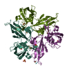
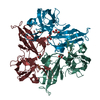
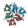
| Title | Two-Domain Laccase from Streptomyces viridochromogenes at 2.4 A resolution AC629 | ||||||
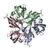
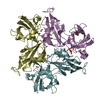
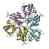
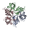
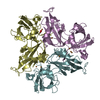
 Components Components | Multicopper oxidase | ||||||
 Keywords Keywords | OXIDOREDUCTASE / TWO-DOMAIN LACCASE / MULTICOPPER BLUE PROTEIN | ||||||
| Function / homology |  Function and homology information Function and homology informationhydroquinone:oxygen oxidoreductase activity / laccase / iron ion transport / copper ion binding / plasma membrane Similarity search - Function | ||||||
| Biological species |  Streptomyces viridochromogenes (bacteria) Streptomyces viridochromogenes (bacteria) | ||||||
| Method |  X-RAY DIFFRACTION / SYNCHROTRON / MOLECULAR REPLACEMENT / Resolution: 2.4 Å X-RAY DIFFRACTION / SYNCHROTRON / MOLECULAR REPLACEMENT / Resolution: 2.4 Å | ||||||
 Authors Authors | Gabdulkhakov, A. / Tischenko, S. / Yurevich, L. / Lisov, A. / Leontievsky, A. | ||||||
 Citation Citation | Journal: To be Published Title: Two-Domain Laccase from Streptomyces viridochromogenes at 2.4 A resolution AC629 Authors: Gabdulkhakov, A. / Tischenko, S. / Yurevich, L. / Lisov, A. / Leontievsky, A. | ||||||
| History |
|
- Structure visualization
| Structure viewer | Molecule: MolmilJmol/JSmol |
|---|
- Downloads & links
-Download
| PDBx/mmCIF format | 4n8u.cif.gz | 170.5 KB | Display | PDBx/mmCIF format |
|---|---|---|---|---|
| PDB format | pdb4n8u.ent.gz | 133.5 KB | Display | PDB format |
| PDBx/mmJSON format | 4n8u.json.gz | Tree view | PDBx/mmJSON format | |
| Others |  Other downloads Other downloads |
-Validation report
| Arichive directory | https://data.pdbj.org/pub/pdb/validation_reports/n8/4n8uftp://data.pdbj.org/pub/pdb/validation_reports/n8/4n8u | HTTPS FTP |
|---|
-Related structure data
| Related structure data | 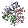 3tasS S: Starting model for refinement |
|---|---|
| Similar structure data |


-Links
 PDBj
PDBj

- Assembly
| Deposited unit | 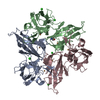
| ||||||||
|---|---|---|---|---|---|---|---|---|---|
| 1 |
| ||||||||
| Unit cell |
|
-Components
| #1: Protein | Mass: 30647.119 Da / Num. of mol.: 3 / Fragment: UNP residues 33-313 Source method: isolated from a genetically manipulated source Source: (gene. exp.) Streptomyces viridochromogenes (bacteria)Plasmid: pQE30 / Production host: #2: Chemical | ChemComp-CU /   Mass: 63.546 Da / Num. of mol.: 9 / Source method: obtained synthetically / Formula: Cu Mass: 63.546 Da / Num. of mol.: 9 / Source method: obtained synthetically / Formula: Cu#3: Chemical | ChemComp-CL /   Mass: 35.453 Da / Num. of mol.: 6 / Source method: obtained synthetically / Formula: Cl Mass: 35.453 Da / Num. of mol.: 6 / Source method: obtained synthetically / Formula: Cl#4: Chemical | ChemComp-TRS / |   Mass: 122.143 Da / Num. of mol.: 1 / Source method: obtained synthetically / Formula: C4H12NO3 / Comment: pH buffer*YM Mass: 122.143 Da / Num. of mol.: 1 / Source method: obtained synthetically / Formula: C4H12NO3 / Comment: pH buffer*YM#5: Water | ChemComp-HOH / |  Mass: 18.015 Da / Num. of mol.: 83 / Source method: isolated from a natural source / Formula: H2O Mass: 18.015 Da / Num. of mol.: 83 / Source method: isolated from a natural source / Formula: H2O |
|---|
-Experimental details
-Experiment
| Experiment | Method: X-RAY DIFFRACTION / Number of used crystals: 1 |
|---|
- Sample preparation
| Crystal | Density Matthews: 2.15 Å3/Da / Density % sol: 42.84 % |
|---|---|
| Crystal grow | Temperature: 285 K / Method: vapor diffusion, hanging drop / pH: 7.5 Details: 11% PEG 4000, 0.1M Tris- l, 0.1M NaCl, 1.3% polyacrylic acid 5100 sodium salt, pH 7.5, VAPOR DIFFUSION, HANGING DROP, temperature 285K |
-Data collection
| Diffraction | Mean temperature: 100 K |
|---|---|
| Diffraction source | Source: SYNCHROTRON / Site: BESSY  / Beamline: 14.1 / Wavelength: 0.91841 Å / Beamline: 14.1 / Wavelength: 0.91841 Å |
| Detector | Type: RAYONIX MX-225 / Detector: CCD / Date: Sep 25, 2012 / Details: mirrors |
| Radiation | Monochromator: Si-111 crystal / Protocol: SINGLE WAVELENGTH / Monochromatic (M) / Laue (L): M / Scattering type: x-ray |
| Radiation wavelength | Wavelength: 0.91841 Å / Relative weight: 1 |
| Reflection | Resolution: 2.4→50 Å / Num. all: 31995 / Num. obs: 31212 / % possible obs: 97.6 % / Observed criterion σ(F): 0 / Observed criterion σ(I): 0 / Redundancy: 3.45 % / Biso Wilson estimate: 20.42 Å2 / Rmerge(I) obs: 0.15 / Net I/σ(I): 7.98 |
| Reflection shell | Resolution: 2.4→2.5 Å / Redundancy: 2.85 % / Rmerge(I) obs: 0.418 / Mean I/σ(I) obs: 2.63 / Num. unique all: 3607 / % possible all: 87.8 |
- Processing
| Software |
| ||||||||||||||||||||||||||||||||||||||||||||||||||||||||||||||||||||||||||||||||||||
|---|---|---|---|---|---|---|---|---|---|---|---|---|---|---|---|---|---|---|---|---|---|---|---|---|---|---|---|---|---|---|---|---|---|---|---|---|---|---|---|---|---|---|---|---|---|---|---|---|---|---|---|---|---|---|---|---|---|---|---|---|---|---|---|---|---|---|---|---|---|---|---|---|---|---|---|---|---|---|---|---|---|---|---|---|---|
| Refinement | Method to determine structure: MOLECULAR REPLACEMENT Starting model: 3TAS Resolution: 2.4→42.95 Å / Occupancy max: 1 / Occupancy min: 1 / FOM work R set: 0.8889 / SU ML: 0.33 / σ(F): 2.05 / Phase error: 20.21 / Stereochemistry target values: ML
| ||||||||||||||||||||||||||||||||||||||||||||||||||||||||||||||||||||||||||||||||||||
| Solvent computation | Shrinkage radii: 0.9 Å / VDW probe radii: 1.11 Å / Solvent model: FLAT BULK SOLVENT MODEL | ||||||||||||||||||||||||||||||||||||||||||||||||||||||||||||||||||||||||||||||||||||
| Displacement parameters | Biso max: 82.36 Å2 / Biso mean: 20.8567 Å2 / Biso min: 4.79 Å2 | ||||||||||||||||||||||||||||||||||||||||||||||||||||||||||||||||||||||||||||||||||||
| Refinement step | Cycle: LAST / Resolution: 2.4→42.95 Å
| ||||||||||||||||||||||||||||||||||||||||||||||||||||||||||||||||||||||||||||||||||||
| Refine LS restraints |
| ||||||||||||||||||||||||||||||||||||||||||||||||||||||||||||||||||||||||||||||||||||
| LS refinement shell | Refine-ID: X-RAY DIFFRACTION / Total num. of bins used: 11
|
