regulation of endoribonuclease activity / TFAP2A acts as a transcriptional repressor during retinoic acid induced cell differentiation / TP53 regulates transcription of additional cell cycle genes whose exact role in the p53 pathway remain uncertain / regulation of endodeoxyribonuclease activity / regulation of eIF2 alpha phosphorylation by dsRNA / positive regulation of cell cycle G2/M phase transition / Deposition of new CENPA-containing nucleosomes at the centromere / SUMOylation of transcription cofactors / regulation of centriole replication / granular component ...regulation of endoribonuclease activity / TFAP2A acts as a transcriptional repressor during retinoic acid induced cell differentiation / TP53 regulates transcription of additional cell cycle genes whose exact role in the p53 pathway remain uncertain / regulation of endodeoxyribonuclease activity / regulation of eIF2 alpha phosphorylation by dsRNA / positive regulation of cell cycle G2/M phase transition / Deposition of new CENPA-containing nucleosomes at the centromere / SUMOylation of transcription cofactors / regulation of centriole replication / granular component / positive regulation of centrosome duplication / positive regulation of protein localization to nucleolus / negative regulation of protein kinase activity by regulation of protein phosphorylation / PKR-mediated signaling / regulation of centrosome duplication / cell volume homeostasis / centrosome cycle / regulation of DNA damage response, signal transduction by p53 class mediator / nucleocytoplasmic transport / post-transcriptional regulation of gene expression / ribosomal large subunit binding / negative regulation of mRNA splicing, via spliceosome / protein kinase inhibitor activity / ribosomal small subunit binding / ribosomal large subunit export from nucleus / NF-kappaB binding / ribosomal small subunit export from nucleus / positive regulation of protein ubiquitination / ribosomal large subunit biogenesis / positive regulation of translation / regulation of protein stability / regulation of cell growth / cellular senescence / : / intracellular protein localization / large ribosomal subunit / ribosomal small subunit biogenesis / small ribosomal subunit / regulation of cell cycle / nuclear speck / rRNA binding / protein stabilization / ribonucleoprotein complex / negative regulation of cell population proliferation / negative regulation of gene expression / DNA repair / positive regulation of cell population proliferation / centrosome / negative regulation of apoptotic process / positive regulation of DNA-templated transcription / nucleolus / positive regulation of transcription by RNA polymerase II / DNA binding / RNA binding / nucleoplasm / identical protein binding / nucleus / cytoplasm / cytosol Similarity search - Function
In the structure databanks used in Yorodumi, some data are registered as the other names, "COVID-19 virus" and "2019-nCoV". Here are the details of the virus and the list of structure data.
Jan 31, 2019. EMDB accession codes are about to change! (news from PDBe EMDB page)
EMDB accession codes are about to change! (news from PDBe EMDB page)
The allocation of 4 digits for EMDB accession codes will soon come to an end. Whilst these codes will remain in use, new EMDB accession codes will include an additional digit and will expand incrementally as the available range of codes is exhausted. The current 4-digit format prefixed with “EMD-” (i.e. EMD-XXXX) will advance to a 5-digit format (i.e. EMD-XXXXX), and so on. It is currently estimated that the 4-digit codes will be depleted around Spring 2019, at which point the 5-digit format will come into force.
The EM Navigator/Yorodumi systems omit the EMD- prefix.
Related info.:Q: What is EMD? / ID/Accession-code notation in Yorodumi/EM Navigator
Yorodumi is a browser for structure data from EMDB, PDB, SASBDB, etc.
This page is also the successor to EM Navigator detail page, and also detail information page/front-end page for Omokage search.
The word "yorodu" (or yorozu) is an old Japanese word meaning "ten thousand". "mi" (miru) is to see.
Related info.:EMDB / PDB / SASBDB / Comparison of 3 databanks / Yorodumi Search / Aug 31, 2016. New EM Navigator & Yorodumi / Yorodumi Papers / Jmol/JSmol / Function and homology information / Changes in new EM Navigator and Yorodumi
 Movie
Movie Controller

 Yorodumi
 Open data
 Basic information
 Components
 Keywords
 Function and homology information
 X-RAY DIFFRACTION /
  Authors
 Citation
 Structure visualization
 Downloads & links
 Other downloads
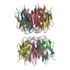
 PDBj
PDBj


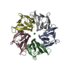
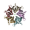
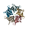
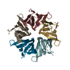
 Assembly
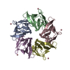


 Mass: 58.933 Da / Num. of mol.: 5 / Source method: obtained synthetically / Formula: Co
 Mass: 18.015 Da / Num. of mol.: 227 / Source method: isolated from a natural source / Formula: H2O
 Sample preparation
 / Beamline: 22-ID / Wavelength: 1 Å
 Processing