 Movie
Movie Controller

[English] 日本語
 Yorodumi
- PDB-4n7v: Crystal structure of human Plk4 cryptic polo box (CPB) in complex... -
+ Open data

- Basic information
| Entry | Database: PDB / ID: 4n7v | ||||||
|---|---|---|---|---|---|---|---|
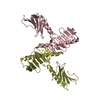
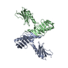
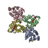
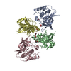
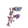
| Title | Crystal structure of human Plk4 cryptic polo box (CPB) in complex with a Cep152 N-terminal fragment | ||||||
 Components Components |
| ||||||
 Keywords Keywords | CELL CYCLE / K/R crater / D-rich motif / Centriole biogenesis / Cep152 / Centrosome | ||||||
| Function / homology |  Function and homology information Function and homology informationde novo centriole assembly involved in multi-ciliated epithelial cell differentiation / procentriole / deuterosome / procentriole replication complex / positive regulation of centriole replication / trophoblast giant cell differentiation / polo kinase / XY body / cell projection organization / pericentriolar material ...de novo centriole assembly involved in multi-ciliated epithelial cell differentiation / procentriole / deuterosome / procentriole replication complex / positive regulation of centriole replication / trophoblast giant cell differentiation / polo kinase / XY body / cell projection organization / pericentriolar material / centriole replication / cleavage furrow / centrosome duplication / cilium assembly / Loss of Nlp from mitotic centrosomes / Loss of proteins required for interphase microtubule organization from the centrosome / Recruitment of mitotic centrosome proteins and complexes / Recruitment of NuMA to mitotic centrosomes / Anchoring of the basal body to the plasma membrane / AURKA Activation by TPX2 / centriole / Regulation of PLK1 Activity at G2/M Transition / protein phosphorylation / nuclear body / ciliary basal body / protein serine kinase activity / protein serine/threonine kinase activity / centrosome / protein kinase binding / nucleolus / nucleoplasm / ATP binding / identical protein binding / nucleus / cytosol Similarity search - Function | ||||||
| Biological species |  Homo sapiens (human) Homo sapiens (human) | ||||||
| Method |  X-RAY DIFFRACTION / SYNCHROTRON / MOLECULAR REPLACEMENT / Resolution: 2.758 Å X-RAY DIFFRACTION / SYNCHROTRON / MOLECULAR REPLACEMENT / Resolution: 2.758 Å | ||||||
 Authors Authors | Park, S.-Y. / Park, J.-E. / Tian, L. / Kim, T.-S. / Yang, W. / Lee, K.S. | ||||||
 Citation Citation | Journal: Nat.Struct.Mol.Biol. / Year: 2014 Title: Molecular basis for unidirectional scaffold switching of human Plk4 in centriole biogenesis. Authors: Park, S.Y. / Park, J.E. / Kim, T.S. / Kim, J.H. / Kwak, M.J. / Ku, B. / Tian, L. / Murugan, R.N. / Ahn, M. / Komiya, S. / Hojo, H. / Kim, N.H. / Kim, B.Y. / Bang, J.K. / Erikson, R.L. / Lee, ...Authors: Park, S.Y. / Park, J.E. / Kim, T.S. / Kim, J.H. / Kwak, M.J. / Ku, B. / Tian, L. / Murugan, R.N. / Ahn, M. / Komiya, S. / Hojo, H. / Kim, N.H. / Kim, B.Y. / Bang, J.K. / Erikson, R.L. / Lee, K.W. / Kim, S.J. / Oh, B.H. / Yang, W. / Lee, K.S. | ||||||
| History |
|
- Structure visualization
| Structure viewer | Molecule: MolmilJmol/JSmol |
|---|
- Downloads & links
-Download
| PDBx/mmCIF format | 4n7v.cif.gz | 109.4 KB | Display | PDBx/mmCIF format |
|---|---|---|---|---|
| PDB format | pdb4n7v.ent.gz | 84.7 KB | Display | PDB format |
| PDBx/mmJSON format | 4n7v.json.gz | Tree view | PDBx/mmJSON format | |
| Others |  Other downloads Other downloads |
-Validation report
| Arichive directory | https://data.pdbj.org/pub/pdb/validation_reports/n7/4n7vftp://data.pdbj.org/pub/pdb/validation_reports/n7/4n7v | HTTPS FTP |
|---|
-Related structure data
| Related structure data | 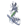 4n7zC 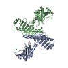 4n9jC  4g7nS C: citing same article ( S: Starting model for refinement |
|---|---|
| Similar structure data |


-Links
 PDBj
PDBj- Assembly
| Deposited unit | 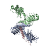
| ||||||||
|---|---|---|---|---|---|---|---|---|---|
| 1 |
| ||||||||
| Unit cell |
|
-Components
| #1: Protein | Mass: 26575.330 Da / Num. of mol.: 2 / Fragment: UNP residues 580-808 Source method: isolated from a genetically manipulated source Source: (gene. exp.) Homo sapiens (human) / Gene: PLK4, SAK, STK18 / Production host:  #2: Protein | | Mass: 6945.263 Da / Num. of mol.: 1 / Fragment: UNP residues 1-60 Source method: isolated from a genetically manipulated source Source: (gene. exp.) Homo sapiens (human) / Gene: CEP152, KIAA0912 / Production host: #3: Water | ChemComp-HOH / |  Mass: 18.015 Da / Num. of mol.: 16 / Source method: isolated from a natural source / Formula: H2O Mass: 18.015 Da / Num. of mol.: 16 / Source method: isolated from a natural source / Formula: H2O |
|---|
-Experimental details
-Experiment
| Experiment | Method: X-RAY DIFFRACTION / Number of used crystals: 1 |
|---|
- Sample preparation
| Crystal | Density Matthews: 2.68 Å3/Da / Density % sol: 54.04 % |
|---|---|
| Crystal grow | Temperature: 295.15 K / Method: vapor diffusion, hanging drop / pH: 8.5 Details: 22% PEG 4000, 3% Iso-propanol, 0.1M Tris-HCl, 0.2M Lithium Sulfate , pH 8.5, VAPOR DIFFUSION, HANGING DROP, temperature 295.15K |
-Data collection
| Diffraction | Mean temperature: 100 K |
|---|---|
| Diffraction source | Source: SYNCHROTRON / Site: APS  / Beamline: 22-BM / Wavelength: 1 Å / Beamline: 22-BM / Wavelength: 1 Å |
| Detector | Type: MARMOSAIC 225 mm CCD / Detector: CCD / Date: Jun 2, 2013 |
| Radiation | Monochromator: double crystal - liqued nitrogen cooled / Protocol: SINGLE WAVELENGTH / Monochromatic (M) / Laue (L): M / Scattering type: x-ray |
| Radiation wavelength | Wavelength: 1 Å / Relative weight: 1 |
| Reflection | Resolution: 2.75→50 Å / Num. obs: 15869 / % possible obs: 92.6 % / Observed criterion σ(F): -0.7 / Observed criterion σ(I): -0.7 / Redundancy: 3.8 % / Rmerge(I) obs: 0.068 / Rsym value: 0.06 / Net I/σ(I): 16.9 |
| Reflection shell | Resolution: 2.75→2.8 Å / Redundancy: 2 % / Rmerge(I) obs: 0.303 / Mean I/σ(I) obs: 2.06 / Num. unique all: 605 / Rsym value: 0.315 / % possible all: 72.7 |
- Processing
| Software |
| ||||||||||||||||||||||||||||||||||||||||||||||||||||||||||||||||||||||||||||||||||||
|---|---|---|---|---|---|---|---|---|---|---|---|---|---|---|---|---|---|---|---|---|---|---|---|---|---|---|---|---|---|---|---|---|---|---|---|---|---|---|---|---|---|---|---|---|---|---|---|---|---|---|---|---|---|---|---|---|---|---|---|---|---|---|---|---|---|---|---|---|---|---|---|---|---|---|---|---|---|---|---|---|---|---|---|---|---|
| Refinement | Method to determine structure: MOLECULAR REPLACEMENT Starting model: PDB ENTRY 4G7N Resolution: 2.758→37.263 Å / SU ML: 0.45 / σ(F): 1.41 / Phase error: 31.55 / Stereochemistry target values: ML
| ||||||||||||||||||||||||||||||||||||||||||||||||||||||||||||||||||||||||||||||||||||
| Solvent computation | Shrinkage radii: 0.9 Å / VDW probe radii: 1.11 Å / Solvent model: FLAT BULK SOLVENT MODEL | ||||||||||||||||||||||||||||||||||||||||||||||||||||||||||||||||||||||||||||||||||||
| Refinement step | Cycle: LAST / Resolution: 2.758→37.263 Å
| ||||||||||||||||||||||||||||||||||||||||||||||||||||||||||||||||||||||||||||||||||||
| Refine LS restraints |
| ||||||||||||||||||||||||||||||||||||||||||||||||||||||||||||||||||||||||||||||||||||
| LS refinement shell |
|
