 Movie
Movie Controller

[English] 日本語
 Yorodumi
- PDB-4n5v: Alternative substrates of Mycobacterium tuberculosis anthranilate... -
+ Open data

- Basic information
| Entry | Database: PDB / ID: 4n5v | ||||||
|---|---|---|---|---|---|---|---|
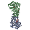
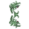
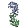
| Title | Alternative substrates of Mycobacterium tuberculosis anthranilate phosphoribosyl transferase | ||||||
 Components Components | Anthranilate phosphoribosyltransferase | ||||||
 Keywords Keywords | TRANSFERASE / Anthranilate Phosphoribosyltransferase / Anthranilic Acids / Magnesium / Tryptophan / Inhibitor / Magnesium binding Phosphoribosyl pyrophosphate | ||||||
| Function / homology |  Function and homology information Function and homology informationanthranilate phosphoribosyltransferase / anthranilate phosphoribosyltransferase activity / L-tryptophan biosynthetic process / magnesium ion binding / extracellular region / plasma membrane / cytosol Similarity search - Function | ||||||
| Biological species |   Mycobacterium tuberculosis (bacteria) Mycobacterium tuberculosis (bacteria) | ||||||
| Method |  X-RAY DIFFRACTION / SYNCHROTRON / MOLECULAR REPLACEMENT / Resolution: 1.9 Å X-RAY DIFFRACTION / SYNCHROTRON / MOLECULAR REPLACEMENT / Resolution: 1.9 Å | ||||||
 Authors Authors | Castell, A. / Cookson, T.V.M. / Parker, E.J. / Baker, E.N. / Lott, J.S. | ||||||
 Citation Citation | Journal: Biochem.J. / Year: 2014 Title: Alternative substrates reveal catalytic cycle and key binding events in the reaction catalysed by anthranilate phosphoribosyltransferase from Mycobacterium tuberculosis. Authors: Cookson, T.V. / Castell, A. / Bulloch, E.M. / Evans, G.L. / Short, F.L. / Baker, E.N. / Lott, J.S. / Parker, E.J. | ||||||
| History |
|
- Structure visualization
| Structure viewer | Molecule: MolmilJmol/JSmol |
|---|
- Downloads & links
-Download
| PDBx/mmCIF format | 4n5v.cif.gz | 153.3 KB | Display | PDBx/mmCIF format |
|---|---|---|---|---|
| PDB format | pdb4n5v.ent.gz | 119.4 KB | Display | PDB format |
| PDBx/mmJSON format | 4n5v.json.gz | Tree view | PDBx/mmJSON format | |
| Others |  Other downloads Other downloads |
-Validation report
| Arichive directory | https://data.pdbj.org/pub/pdb/validation_reports/n5/4n5vftp://data.pdbj.org/pub/pdb/validation_reports/n5/4n5v | HTTPS FTP |
|---|
-Related structure data
| Related structure data |  4n8qC  4n93C  4owmC  4ownC  4owoC  4owqC  4owsC  4owuC  4owvC  3qr9S S: Starting model for refinement C: citing same article ( |
|---|---|
| Similar structure data |


-Links
 PDBj
PDBj
- Assembly
| Deposited unit | 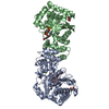
| ||||||||
|---|---|---|---|---|---|---|---|---|---|
| 1 |
| ||||||||
| Unit cell |
|
-Components
-Protein / Sugars , 2 types, 4 molecules AB
| #1: Protein | Mass: 38790.848 Da / Num. of mol.: 2 Source method: isolated from a genetically manipulated source Source: (gene. exp.) Mycobacterium tuberculosis (bacteria) / Strain: H37Rv / Gene: MT2248, MTCY190.03c, Rv2192c, trpD / Plasmid: pET23a / Production host: References: UniProt: P66992, UniProt: P9WFX5*PLUS, anthranilate phosphoribosyltransferase #2: Sugar |  Type: D-saccharide / Mass: 390.070 Da / Num. of mol.: 2 / Source method: obtained synthetically / Formula: C5H13O14P3 Type: D-saccharide / Mass: 390.070 Da / Num. of mol.: 2 / Source method: obtained synthetically / Formula: C5H13O14P3 |
|---|
-Non-polymers , 4 types, 524 molecules 


| #3: Chemical | ChemComp-MG /  Mass: 24.305 Da / Num. of mol.: 4 / Source method: obtained synthetically / Formula: Mg Mass: 24.305 Da / Num. of mol.: 4 / Source method: obtained synthetically / Formula: Mg#4: Chemical | ChemComp-FA0 /  Mass: 155.126 Da / Num. of mol.: 4 / Source method: obtained synthetically / Formula: C7H6FNO2 Mass: 155.126 Da / Num. of mol.: 4 / Source method: obtained synthetically / Formula: C7H6FNO2#5: Chemical |  Mass: 92.094 Da / Num. of mol.: 3 / Source method: obtained synthetically / Formula: C3H8O3 Mass: 92.094 Da / Num. of mol.: 3 / Source method: obtained synthetically / Formula: C3H8O3#6: Water | ChemComp-HOH / | Mass: 18.015 Da / Num. of mol.: 513 / Source method: isolated from a natural source / Formula: H2O |
|---|
-Experimental details
-Experiment
| Experiment | Method: X-RAY DIFFRACTION / Number of used crystals: 1 |
|---|
- Sample preparation
| Crystal | Density Matthews: 2.87 Å3/Da / Density % sol: 57.2 % |
|---|---|
| Crystal grow | Temperature: 296 K / Method: vapor diffusion, hanging drop / pH: 8 Details: 0.1 M imidazole/malate, 7.5% PEG4000, pH 8.0, VAPOR DIFFUSION, HANGING DROP, temperature 296K |
-Data collection
| Diffraction | Mean temperature: 100 K |
|---|---|
| Diffraction source | Source: SYNCHROTRON / Site: Australian Synchrotron  / Beamline: MX2 / Wavelength: 0.95365 Å / Beamline: MX2 / Wavelength: 0.95365 Å |
| Detector | Type: ADSC QUANTUM 315r / Detector: CCD / Date: Feb 13, 2010 |
| Radiation | Monochromator: Silicon double crystal / Protocol: SINGLE WAVELENGTH / Monochromatic (M) / Laue (L): M / Scattering type: x-ray |
| Radiation wavelength | Wavelength: 0.95365 Å / Relative weight: 1 |
| Reflection | Resolution: 1.9→121.06 Å / Num. obs: 71154 / % possible obs: 100 % / Observed criterion σ(F): 0 / Observed criterion σ(I): -3 / Redundancy: 7 % / Biso Wilson estimate: 25.4 Å2 / Rmerge(I) obs: 0.1 / Rsym value: 0.108 / Net I/σ(I): 14.7 |
| Reflection shell | Resolution: 1.9→2 Å / Redundancy: 6.8 % / Rmerge(I) obs: 1.009 / Mean I/σ(I) obs: 2 / Num. unique all: 69643 / Rsym value: 1.093 / % possible all: 100 |
- Processing
| Software |
| |||||||||||||||||||||||||||||||||||||||||||||||||||||||||||||||||
|---|---|---|---|---|---|---|---|---|---|---|---|---|---|---|---|---|---|---|---|---|---|---|---|---|---|---|---|---|---|---|---|---|---|---|---|---|---|---|---|---|---|---|---|---|---|---|---|---|---|---|---|---|---|---|---|---|---|---|---|---|---|---|---|---|---|---|
| Refinement | Method to determine structure: MOLECULAR REPLACEMENT Starting model: PDB ENTRY 3QR9 Resolution: 1.9→73.52 Å / Cor.coef. Fo:Fc: 0.96 / Cor.coef. Fo:Fc free: 0.944 / SU B: 3.026 / SU ML: 0.088 / Cross valid method: THROUGHOUT / σ(I): 2 / ESU R: 0.126 / ESU R Free: 0.123 / Stereochemistry target values: MAXIMUM LIKELIHOOD / Details: HYDROGENS HAVE BEEN ADDED IN THE RIDING POSITIONS
| |||||||||||||||||||||||||||||||||||||||||||||||||||||||||||||||||
| Solvent computation | Ion probe radii: 0.8 Å / Shrinkage radii: 0.8 Å / VDW probe radii: 1.4 Å / Solvent model: MASK | |||||||||||||||||||||||||||||||||||||||||||||||||||||||||||||||||
| Displacement parameters | Biso mean: 26.619 Å2
| |||||||||||||||||||||||||||||||||||||||||||||||||||||||||||||||||
| Refinement step | Cycle: LAST / Resolution: 1.9→73.52 Å
| |||||||||||||||||||||||||||||||||||||||||||||||||||||||||||||||||
| Refine LS restraints |
| |||||||||||||||||||||||||||||||||||||||||||||||||||||||||||||||||
| LS refinement shell | Resolution: 1.9→1.949 Å / Total num. of bins used: 20
|
