 Movie
Movie Controller

[English] 日本語
 Yorodumi
- PDB-4x5c: Anthranilate phosphoribosyltransferase variant R193L from Mycobac... -
+ Open data

- Basic information
| Entry | Database: PDB / ID: 4x5c | ||||||
|---|---|---|---|---|---|---|---|
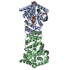
| Title | Anthranilate phosphoribosyltransferase variant R193L from Mycobacterium tuberculosis with pyrophosphate/PRPP and Mg2+ bound | ||||||
 Components Components | Anthranilate phosphoribosyltransferase | ||||||
 Keywords Keywords | TRANSFERASE / Anthranilate Phosphoribosyltransferase / Anthranilic Acids / Mycobacterium tuberculosis / Magnesium / Tryptophan / mutation / Magnesium binding / Phosphoribosyl pyrophosphate | ||||||
| Function / homology |  Function and homology information Function and homology informationanthranilate phosphoribosyltransferase / anthranilate phosphoribosyltransferase activity / L-tryptophan biosynthetic process / magnesium ion binding / extracellular region / plasma membrane / cytosol Similarity search - Function | ||||||
| Biological species |   Mycobacterium tuberculosis (bacteria) Mycobacterium tuberculosis (bacteria) | ||||||
| Method |  X-RAY DIFFRACTION / SYNCHROTRON / MOLECULAR REPLACEMENT / Resolution: 2.33 Å X-RAY DIFFRACTION / SYNCHROTRON / MOLECULAR REPLACEMENT / Resolution: 2.33 Å | ||||||
 Authors Authors | Cookson, T.V.M. / Parker, E.J. / Lott, J.S. | ||||||
 Citation Citation | Journal: Biochemistry / Year: 2015 Title: Structures of Mycobacterium tuberculosis Anthranilate Phosphoribosyltransferase Variants Reveal the Conformational Changes That Facilitate Delivery of the Substrate to the Active Site. Authors: Cookson, T.V. / Evans, G.L. / Castell, A. / Baker, E.N. / Lott, J.S. / Parker, E.J. | ||||||
| History |
|
- Structure visualization
| Structure viewer | Molecule: MolmilJmol/JSmol |
|---|
- Downloads & links
-Download
| PDBx/mmCIF format | 4x5c.cif.gz | 256.7 KB | Display | PDBx/mmCIF format |
|---|---|---|---|---|
| PDB format | pdb4x5c.ent.gz | 206.7 KB | Display | PDB format |
| PDBx/mmJSON format | 4x5c.json.gz | Tree view | PDBx/mmJSON format | |
| Others |  Other downloads Other downloads |
-Validation report
| Arichive directory | https://data.pdbj.org/pub/pdb/validation_reports/x5/4x5cftp://data.pdbj.org/pub/pdb/validation_reports/x5/4x5c | HTTPS FTP |
|---|
-Related structure data
| Related structure data |  4x58C  4x59C  4x5aC  4x5bC  4x5dC  4x5eC  3qr9S C: citing same article ( S: Starting model for refinement |
|---|---|
| Similar structure data |


-Links
 PDBj
PDBj


- Assembly
| Deposited unit | 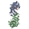
| ||||||||
|---|---|---|---|---|---|---|---|---|---|
| 1 |
| ||||||||
| Unit cell |
| ||||||||
| Components on special symmetry positions |
|
-Components
| #1: Protein | Mass: 38804.848 Da / Num. of mol.: 2 / Mutation: R193L Source method: isolated from a genetically manipulated source Source: (gene. exp.) Mycobacterium tuberculosis (bacteria) / Gene: trpD / Plasmid: pET23a / Production host: References: UniProt: A5U4M0, UniProt: P9WFX5*PLUS, anthranilate phosphoribosyltransferase #2: Sugar | ChemComp-PRP / |   Type: D-saccharide / Mass: 390.070 Da / Num. of mol.: 1 / Source method: obtained synthetically / Formula: C5H13O14P3 Type: D-saccharide / Mass: 390.070 Da / Num. of mol.: 1 / Source method: obtained synthetically / Formula: C5H13O14P3#3: Chemical | ChemComp-MG /   Mass: 24.305 Da / Num. of mol.: 4 / Source method: obtained synthetically / Formula: Mg Mass: 24.305 Da / Num. of mol.: 4 / Source method: obtained synthetically / Formula: Mg#4: Chemical | ChemComp-POP / |   Mass: 175.959 Da / Num. of mol.: 1 / Source method: obtained synthetically / Formula: H2O7P2 Mass: 175.959 Da / Num. of mol.: 1 / Source method: obtained synthetically / Formula: H2O7P2#5: Water | ChemComp-HOH / |  Mass: 18.015 Da / Num. of mol.: 61 / Source method: isolated from a natural source / Formula: H2O Mass: 18.015 Da / Num. of mol.: 61 / Source method: isolated from a natural source / Formula: H2O |
|---|
-Experimental details
-Experiment
| Experiment | Method: X-RAY DIFFRACTION / Number of used crystals: 1 |
|---|
- Sample preparation
| Crystal | Density Matthews: 2.43 Å3/Da / Density % sol: 49.45 % |
|---|---|
| Crystal grow | Temperature: 293 K / Method: vapor diffusion, hanging drop / Details: imidazole malate, PEG 4000 |
-Data collection
| Diffraction | Mean temperature: 110 K | |||||||||||||||||||||||||||
|---|---|---|---|---|---|---|---|---|---|---|---|---|---|---|---|---|---|---|---|---|---|---|---|---|---|---|---|---|
| Diffraction source | Source: SYNCHROTRON / Site: Australian Synchrotron  / Beamline: MX1 / Wavelength: 0.9537 Å / Beamline: MX1 / Wavelength: 0.9537 Å | |||||||||||||||||||||||||||
| Detector | Type: ADSC QUANTUM 210r / Detector: CCD / Date: Dec 3, 2012 | |||||||||||||||||||||||||||
| Radiation | Protocol: SINGLE WAVELENGTH / Monochromatic (M) / Laue (L): M / Scattering type: x-ray | |||||||||||||||||||||||||||
| Radiation wavelength | Wavelength: 0.9537 Å / Relative weight: 1 | |||||||||||||||||||||||||||
| Reflection | Resolution: 2.33→94.12 Å / Num. obs: 29405 / % possible obs: 100 % / Redundancy: 7.4 % / CC1/2: 0.996 / Rmerge(I) obs: 0.182 / Rpim(I) all: 0.072 / Net I/σ(I): 10.2 / Num. measured all: 216139 / Scaling rejects: 2 | |||||||||||||||||||||||||||
| Reflection shell | Diffraction-ID: 1 / Rejects: _
|
- Processing
| Software |
| |||||||||||||||||||||||||||||||||||||||||||||||||||||||||||||||||||||||||||
|---|---|---|---|---|---|---|---|---|---|---|---|---|---|---|---|---|---|---|---|---|---|---|---|---|---|---|---|---|---|---|---|---|---|---|---|---|---|---|---|---|---|---|---|---|---|---|---|---|---|---|---|---|---|---|---|---|---|---|---|---|---|---|---|---|---|---|---|---|---|---|---|---|---|---|---|---|
| Refinement | Method to determine structure: MOLECULAR REPLACEMENT Starting model: 3QR9 Resolution: 2.33→94.12 Å / Cor.coef. Fo:Fc: 0.953 / Cor.coef. Fo:Fc free: 0.917 / SU B: 19.716 / SU ML: 0.205 / Cross valid method: THROUGHOUT / σ(F): 0 / ESU R: 0.376 / ESU R Free: 0.258 / Stereochemistry target values: MAXIMUM LIKELIHOOD Details: HYDROGENS HAVE BEEN ADDED IN THE RIDING POSITIONS U VALUES : WITH TLS ADDED
| |||||||||||||||||||||||||||||||||||||||||||||||||||||||||||||||||||||||||||
| Solvent computation | Ion probe radii: 0.8 Å / Shrinkage radii: 0.8 Å / VDW probe radii: 1.2 Å / Solvent model: MASK | |||||||||||||||||||||||||||||||||||||||||||||||||||||||||||||||||||||||||||
| Displacement parameters | Biso max: 89.03 Å2 / Biso mean: 40.194 Å2 / Biso min: 19.3 Å2
| |||||||||||||||||||||||||||||||||||||||||||||||||||||||||||||||||||||||||||
| Refinement step | Cycle: final / Resolution: 2.33→94.12 Å
| |||||||||||||||||||||||||||||||||||||||||||||||||||||||||||||||||||||||||||
| Refine LS restraints |
| |||||||||||||||||||||||||||||||||||||||||||||||||||||||||||||||||||||||||||
| LS refinement shell | Resolution: 2.33→2.391 Å / Total num. of bins used: 20
| |||||||||||||||||||||||||||||||||||||||||||||||||||||||||||||||||||||||||||
| Refinement TLS params. | Method: refined / Refine-ID: X-RAY DIFFRACTION
| |||||||||||||||||||||||||||||||||||||||||||||||||||||||||||||||||||||||||||
| Refinement TLS group |
|
