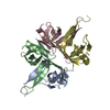
[English] 日本語
Yorodumi
- PDB-4mz9: Revised structure of E. coli SSB -

+
Open data


ID or keywords:

Loading...

-
Basic information

Entry
Database: PDB / ID: 4mz9
TitleRevised structure of E. coli SSB
ComponentsSingle-stranded DNA-binding protein
KeywordsDNA BINDING PROTEIN / Single strand DNA-binding domain / SSB / RecO / ExoI / RecQ / DnaG / HolC
Function / homology
Function and homology information


single-stranded DNA-binding protein complex / nucleoid / replisome / recombinational repair / positive regulation of catalytic activity / SOS response / mismatch repair / enzyme activator activity / DNA-templated DNA replication / single-stranded DNA binding ...single-stranded DNA-binding protein complex / nucleoid / replisome / recombinational repair / positive regulation of catalytic activity / SOS response / mismatch repair / enzyme activator activity / DNA-templated DNA replication / single-stranded DNA binding / identical protein binding / cytosol
Similarity search - Function
Single-stranded DNA-binding protein / Single-strand binding protein family / Single-strand binding (SSB) domain profile. / Primosome PriB/single-strand DNA-binding / Nucleic acid-binding proteins / OB fold (Dihydrolipoamide Acetyltransferase, E2P) / Nucleic acid-binding, OB-fold / Beta Barrel / Mainly Beta
Similarity search - Domain/homology
Single-stranded DNA-binding protein
Similarity search - Component
Biological speciesEscherichia coli (E. coli)
MethodX-RAY DIFFRACTION / MOLECULAR REPLACEMENT / Resolution: 2.2 Å
AuthorsOakley, A.J.
CitationJournal: J.Biochem. / Year: 2000
Title: Roles of functional loops and the C-terminal segment of a single-stranded DNA binding protein elucidated by X-Ray structure analysis.
Authors: Matsumoto, T. / Morimoto, Y. / Shibata, N. / Kinebuchi, T. / Shimamoto, N. / Tsukihara, T. / Yasuoka, N.
History
DepositionSep 29, 2013Deposition site: RCSB / Processing site: RCSB
Revision 1.0Dec 18, 2013Provider: repository / Type: Initial release
Revision 1.1Mar 12, 2014Group: Database references
Revision 1.2Sep 20, 2023Group: Data collection / Database references / Refinement description
Category: chem_comp_atom / chem_comp_bond ...chem_comp_atom / chem_comp_bond / database_2 / pdbx_initial_refinement_model / struct_ncs_dom_lim
Item: _database_2.pdbx_DOI / _database_2.pdbx_database_accession ..._database_2.pdbx_DOI / _database_2.pdbx_database_accession / _struct_ncs_dom_lim.beg_auth_comp_id / _struct_ncs_dom_lim.beg_label_asym_id / _struct_ncs_dom_lim.beg_label_comp_id / _struct_ncs_dom_lim.beg_label_seq_id / _struct_ncs_dom_lim.end_auth_comp_id / _struct_ncs_dom_lim.end_label_asym_id / _struct_ncs_dom_lim.end_label_comp_id / _struct_ncs_dom_lim.end_label_seq_id
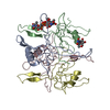
Remark 0THIS ENTRY 4MZ9 REFLECTS AN ALTERNATIVE MODELING OF THE ORIGINAL STRUCTURAL DATA (R1QVCSF) ...THIS ENTRY 4MZ9 REFLECTS AN ALTERNATIVE MODELING OF THE ORIGINAL STRUCTURAL DATA (R1QVCSF) DETERMINED BY AUTHORS OF THE PDB ENTRY 1QVC: T.MATSUMOTO, Y.MORIMOTO, N.SHIBATA, N.SHIMAMOTO, T.TSUKIHARA, N.YASUOKA

-
Structure visualization

Structure viewerMolecule:
MolmilJmol/JSmol

Downloads & links

-
Assembly

Deposited unit
A: Single-stranded DNA-binding protein
B: Single-stranded DNA-binding protein
C: Single-stranded DNA-binding protein
D: Single-stranded DNA-binding protein


Theoretical massNumber of molelcules
Total (without water)75,9684
Polymers75,9684
Non-polymers00
Water2,612145
1


  • Idetical with deposited unit
  • defined by author&software
TypeNameSymmetry operationNumber
identity operation1_555x,y,z1
Buried area7330 Å2
ΔGint-12 kcal/mol
Surface area25740 Å2
MethodPISA
Unit cell
Length a, b, c (Å)105.369, 62.923, 97.792
Angle α, β, γ (deg.)90.00, 112.57, 90.00
Int Tables number5
Space group name H-MC121
Noncrystallographic symmetry (NCS)NCS domain:
IDEns-IDDetails
11A
21B
31C
41D

NCS domain segments:

Ens-ID: 1 / Refine code: 4

Dom-IDComponent-IDBeg auth comp-IDBeg label comp-IDEnd auth comp-IDEnd label comp-IDAuth asym-IDLabel asym-IDAuth seq-IDLabel seq-ID
11ALAALATYRTYRAA1 - 222 - 23
21ALAALATYRTYRBB1 - 222 - 23
31ALAALATYRTYRCC1 - 222 - 23
41ALAALATYRTYRDD1 - 222 - 23
12VALVALSERSERAA29 - 3930 - 40
22VALVALSERSERBB29 - 3930 - 40
32VALVALSERSERCC29 - 3930 - 40
42VALVALSERSERDD29 - 3930 - 40
13METMETHISHISAA48 - 5549 - 56
23METMETHISHISBB48 - 5549 - 56
33METMETHISHISCC48 - 5549 - 56
43METMETHISHISDD48 - 5549 - 56
14VALVALLEULEUAA57 - 8358 - 84
24VALVALLEULEUBB57 - 8358 - 84
34VALVALLEULEUCC57 - 8358 - 84
44VALVALLEULEUDD57 - 8358 - 84
15THRTHRGLNGLNAA85 - 11086 - 111
25THRTHRGLNGLNBB85 - 11086 - 111
35THRTHRGLNGLNCC85 - 11086 - 111
45THRTHRGLNGLNDD85 - 11086 - 111

-
Components

#1: Protein
Single-stranded DNA-binding protein / SSB / Helix-destabilizing protein


Mass: 18991.939 Da / Num. of mol.: 4
Source method: isolated from a genetically manipulated source
Source: (gene. exp.) Escherichia coli (E. coli) / Strain: K12 / Gene: b4059, exrB, JW4020, lexC, ssb / Plasmid: PT7-7 / Production host: Escherichia coli (E. coli) / References: UniProt: P0AGE0
#2: Water ChemComp-HOH / water


Mass: 18.015 Da / Num. of mol.: 145 / Source method: isolated from a natural source / Formula: H2O

-
Experimental details

-
Experiment

ExperimentMethod: X-RAY DIFFRACTION

-
Sample preparation

CrystalDensity Matthews: 1.97 Å3/Da / Density % sol: 37.57 % / Description: AUTHOR USED THE SF DATA FROM ENTRY 1QVC.

-
Data collection

RadiationScattering type: x-ray
Radiation wavelengthRelative weight: 1

-
Processing

SoftwareName: REFMAC / Version: 5.5.0109 / Classification: refinement
RefinementMethod to determine structure: MOLECULAR REPLACEMENT
Starting model: PDB ENTRY 1QVC
Resolution: 2.2→51.958 Å / Cor.coef. Fo:Fc: 0.947 / Cor.coef. Fo:Fc free: 0.908 / SU B: 18.423 / SU ML: 0.207 / Cross valid method: THROUGHOUT / σ(F): 0 / ESU R: 0.322 / ESU R Free: 0.247 / Stereochemistry target values: MAXIMUM LIKELIHOOD / Details: HYDROGENS HAVE BEEN ADDED IN THE RIDING POSITIONS
RfactorNum. reflection% reflectionSelection details
Rfree0.26223 1263 5.1 %RANDOM
Rwork0.20999 ---
all0.21267 24955 --
obs0.21267 23692 82.65 %-
Solvent computationIon probe radii: 0.8 Å / Shrinkage radii: 0.8 Å / VDW probe radii: 1.4 Å / Solvent model: MASK
Displacement parametersBiso mean: 71.1 Å2
Baniso -1Baniso -2Baniso -3
1--1.72 Å20 Å20.4 Å2
2--2.59 Å2-0 Å2
3----0.56 Å2
Refinement stepCycle: LAST / Resolution: 2.2→51.958 Å
ProteinNucleic acidLigandSolventTotal
Num. atoms3536 0 0 145 3681
Refine LS restraints
Refine-IDTypeDev idealDev ideal targetNumber
X-RAY DIFFRACTIONr_bond_refined_d0.020.0223600
X-RAY DIFFRACTIONr_bond_other_d0.0040.022452
X-RAY DIFFRACTIONr_angle_refined_deg1.9581.9424859
X-RAY DIFFRACTIONr_angle_other_deg1.1135951
X-RAY DIFFRACTIONr_dihedral_angle_1_deg9.4485456
X-RAY DIFFRACTIONr_dihedral_angle_2_deg39.20924.083169
X-RAY DIFFRACTIONr_dihedral_angle_3_deg23.49915644
X-RAY DIFFRACTIONr_dihedral_angle_4_deg19.4071532
X-RAY DIFFRACTIONr_chiral_restr0.1380.2534
X-RAY DIFFRACTIONr_gen_planes_refined0.0070.024050
X-RAY DIFFRACTIONr_gen_planes_other0.0010.02726
X-RAY DIFFRACTIONr_nbd_refined
X-RAY DIFFRACTIONr_nbd_other
X-RAY DIFFRACTIONr_nbtor_refined
X-RAY DIFFRACTIONr_nbtor_other
X-RAY DIFFRACTIONr_xyhbond_nbd_refined
X-RAY DIFFRACTIONr_xyhbond_nbd_other
X-RAY DIFFRACTIONr_metal_ion_refined
X-RAY DIFFRACTIONr_metal_ion_other
X-RAY DIFFRACTIONr_symmetry_vdw_refined
X-RAY DIFFRACTIONr_symmetry_vdw_other
X-RAY DIFFRACTIONr_symmetry_hbond_refined
X-RAY DIFFRACTIONr_symmetry_hbond_other
X-RAY DIFFRACTIONr_symmetry_metal_ion_refined
X-RAY DIFFRACTIONr_symmetry_metal_ion_other
X-RAY DIFFRACTIONr_mcbond_it1.10622234
X-RAY DIFFRACTIONr_mcbond_other0.3562958
X-RAY DIFFRACTIONr_mcangle_it1.9233576
X-RAY DIFFRACTIONr_mcangle_other
X-RAY DIFFRACTIONr_scbond_it2.25731366
X-RAY DIFFRACTIONr_scbond_other
X-RAY DIFFRACTIONr_scangle_it3.4151281
X-RAY DIFFRACTIONr_scangle_other
X-RAY DIFFRACTIONr_long_range_B_refined
X-RAY DIFFRACTIONr_long_range_B_other
X-RAY DIFFRACTIONr_rigid_bond_restr
X-RAY DIFFRACTIONr_sphericity_free
X-RAY DIFFRACTIONr_sphericity_bonded
Refine LS restraints NCS

Dom-ID: 1 / Ens-ID: 1 / Number: 1219 / Refine-ID: X-RAY DIFFRACTION

Auth asym-IDTypeRms dev position (Å)Weight position
Amedium positional0.570.5
Bmedium positional0.60.5
Cmedium positional0.540.5
Dmedium positional0.530.5
Amedium thermal0.92
Bmedium thermal0.842
Cmedium thermal0.942
Dmedium thermal0.812
LS refinement shellResolution: 2.2→2.257 Å / Total num. of bins used: 20
RfactorNum. reflection% reflection
Rfree0.2 26 -
Rwork0.251 723 -
obs--34.14 %
Refinement TLS params.

Method: refined / Refine-ID: X-RAY DIFFRACTION

IDL112)L122)L132)L222)L232)L332)S11 (Å °)S12 (Å °)S13 (Å °)S21 (Å °)S22 (Å °)S23 (Å °)S31 (Å °)S32 (Å °)S33 (Å °)T112)T122)T132)T222)T232)T332)Origin x (Å)Origin y (Å)Origin z (Å)
17.68560.88291.55444.2230.46228.120.1942-0.79020.28960.4904-0.24340.5685-0.1274-0.71110.04920.20730.14720.07340.3315-0.04980.236222.99620.148931.9987
25.9997-2.1102-1.14684.31120.87016.88880.06860.5603-0.1124-0.44890.04050.530.1425-0.3971-0.1090.18640.1518-0.09440.35290.0240.282719.628-0.133513.8562
36.95230.25662.16814.49340.88665.3914-0.00260.2684-0.6044-0.46350.06-0.36990.43240.1997-0.05740.27740.20770.05560.2493-0.03090.243645.3186-12.728312.634
46.46750.888-0.85796.67551.30478.57050.2085-0.3410.14110.1057-0.1304-0.7179-0.43411.0886-0.07810.13790.0852-0.00870.3794-0.02480.298750.50331.290923.0493
Refinement TLS group
IDRefine-IDRefine TLS-IDAuth asym-IDAuth seq-ID
1X-RAY DIFFRACTION1A1 - 114
2X-RAY DIFFRACTION1A201 - 230
3X-RAY DIFFRACTION2B1 - 114
4X-RAY DIFFRACTION2B201 - 245
5X-RAY DIFFRACTION3C1 - 114
6X-RAY DIFFRACTION3C201 - 242
7X-RAY DIFFRACTION4D1 - 114
8X-RAY DIFFRACTION4D201 - 228

+
About Yorodumi

-
News

-
Feb 9, 2022. New format data for meta-information of EMDB entries

New format data for meta-information of EMDB entries

  • Version 3 of the EMDB header file is now the official format.
  • The previous official version 1.9 will be removed from the archive.

Related info.:EMDB header

External links:wwPDB to switch to version 3 of the EMDB data model

-
Aug 12, 2020. Covid-19 info

Covid-19 info

URL: https://pdbjlvh1.pdbj.org/emnavi/covid19.php

New page: Covid-19 featured information page in EM Navigator.

Related info.:Covid-19 info / Mar 5, 2020. Novel coronavirus structure data

+
Mar 5, 2020. Novel coronavirus structure data

Novel coronavirus structure data

Related info.:Yorodumi Speices / Aug 12, 2020. Covid-19 info

External links:COVID-19 featured content - PDBj / Molecule of the Month (242):Coronavirus Proteases

+
Jan 31, 2019. EMDB accession codes are about to change! (news from PDBe EMDB page)

EMDB accession codes are about to change! (news from PDBe EMDB page)

  • The allocation of 4 digits for EMDB accession codes will soon come to an end. Whilst these codes will remain in use, new EMDB accession codes will include an additional digit and will expand incrementally as the available range of codes is exhausted. The current 4-digit format prefixed with “EMD-” (i.e. EMD-XXXX) will advance to a 5-digit format (i.e. EMD-XXXXX), and so on. It is currently estimated that the 4-digit codes will be depleted around Spring 2019, at which point the 5-digit format will come into force.
  • The EM Navigator/Yorodumi systems omit the EMD- prefix.

Related info.:Q: What is EMD? / ID/Accession-code notation in Yorodumi/EM Navigator

External links:EMDB Accession Codes are Changing Soon! / Contact to PDBj

+
Jul 12, 2017. Major update of PDB

Major update of PDB

  • wwPDB released updated PDB data conforming to the new PDBx/mmCIF dictionary.
  • This is a major update changing the version number from 4 to 5, and with Remediation, in which all the entries are updated.
  • In this update, many items about electron microscopy experimental information are reorganized (e.g. em_software).
  • Now, EM Navigator and Yorodumi are based on the updated data.

External links:wwPDB Remediation / Enriched Model Files Conforming to OneDep Data Standards Now Available in the PDB FTP Archive

-
Yorodumi

Thousand views of thousand structures

  • Yorodumi is a browser for structure data from EMDB, PDB, SASBDB, etc.
  • This page is also the successor to EM Navigator detail page, and also detail information page/front-end page for Omokage search.
  • The word "yorodu" (or yorozu) is an old Japanese word meaning "ten thousand". "mi" (miru) is to see.

Related info.:EMDB / PDB / SASBDB / Comparison of 3 databanks / Yorodumi Search / Aug 31, 2016. New EM Navigator & Yorodumi / Yorodumi Papers / Jmol/JSmol / Function and homology information / Changes in new EM Navigator and Yorodumi

Read more