 Movie
Movie Controller

[English] 日本語
 Yorodumi
- PDB-4mz0: Structure of a ketosynthase-acyltransferase di-domain from module... -
+ Open data

- Basic information
| Entry | Database: PDB / ID: 4mz0 | ||||||
|---|---|---|---|---|---|---|---|
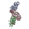
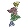
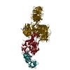
| Title | Structure of a ketosynthase-acyltransferase di-domain from module CurL of the curacin A polyketide synthase | ||||||
 Components Components | CurL | ||||||
 Keywords Keywords | TRANSFERASE / ketosynthase / thiolase fold / acyltransferase / alpha/beta hydrolase fold / Extension of polyketide intermediate | ||||||
| Function / homology |  Function and homology information Function and homology informationDIM/DIP cell wall layer assembly / fatty acid synthase activity / phosphopantetheine binding / 3-oxoacyl-[acyl-carrier-protein] synthase activity / fatty acid biosynthetic process / metal ion binding / plasma membrane / cytoplasm Similarity search - Function | ||||||
| Biological species |  Moorea producens 3L (bacteria) Moorea producens 3L (bacteria) | ||||||
| Method |  X-RAY DIFFRACTION / SYNCHROTRON / MOLECULAR REPLACEMENT / Resolution: 2.8 Å X-RAY DIFFRACTION / SYNCHROTRON / MOLECULAR REPLACEMENT / Resolution: 2.8 Å | ||||||
 Authors Authors | Whicher, J.R. / Smaga, S.S. / Smith, J.L. | ||||||
 Citation Citation | Journal: Chem.Biol. / Year: 2013 Title: Cyanobacterial polyketide synthase docking domains: a tool for engineering natural product biosynthesis. Authors: Whicher, J.R. / Smaga, S.S. / Hansen, D.A. / Brown, W.C. / Gerwick, W.H. / Sherman, D.H. / Smith, J.L. | ||||||
| History |
|
- Structure visualization
| Structure viewer | Molecule: MolmilJmol/JSmol |
|---|
- Downloads & links
-Download
| PDBx/mmCIF format | 4mz0.cif.gz | 340.3 KB | Display | PDBx/mmCIF format |
|---|---|---|---|---|
| PDB format | pdb4mz0.ent.gz | 273.5 KB | Display | PDB format |
| PDBx/mmJSON format | 4mz0.json.gz | Tree view | PDBx/mmJSON format | |
| Others |  Other downloads Other downloads |
-Validation report
| Summary document | 4mz0_validation.pdf.gz | 444.8 KB | Display | wwPDB validaton report |
|---|---|---|---|---|
| Full document | 4mz0_full_validation.pdf.gz | 467.4 KB | Display | |
| Data in XML | 4mz0_validation.xml.gz | 59.5 KB | Display | |
| Data in CIF | 4mz0_validation.cif.gz | 83.1 KB | Display | |
| Arichive directory | https://data.pdbj.org/pub/pdb/validation_reports/mz/4mz0ftp://data.pdbj.org/pub/pdb/validation_reports/mz/4mz0 | HTTPS FTP |
-Related structure data
| Related structure data | 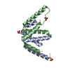 4myyC  4myzC  2qo3S C: citing same article ( S: Starting model for refinement |
|---|---|
| Similar structure data |


-Links
 PDBj
PDBj


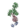
- Assembly
| Deposited unit | 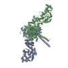
| ||||||||
|---|---|---|---|---|---|---|---|---|---|
| 1 |
| ||||||||
| Unit cell |
|
-Components
| #1: Protein | Mass: 102035.031 Da / Num. of mol.: 2 / Fragment: UNP residues 1-938 Source method: isolated from a genetically manipulated source Source: (gene. exp.) Moorea producens 3L (bacteria) / Gene: LYNGBM3L_74440 / Plasmid: pSUMO / Production host: Strain (production host): BL21 AI containing the pRARE2-CDF plasmid References: UniProt: F4Y424, beta-ketoacyl-[acyl-carrier-protein] synthase I #2: Chemical |   Mass: 40.078 Da / Num. of mol.: 2 / Source method: obtained synthetically / Formula: Ca Mass: 40.078 Da / Num. of mol.: 2 / Source method: obtained synthetically / Formula: Ca#3: Water | ChemComp-HOH / |  Mass: 18.015 Da / Num. of mol.: 282 / Source method: isolated from a natural source / Formula: H2O Mass: 18.015 Da / Num. of mol.: 282 / Source method: isolated from a natural source / Formula: H2O |
|---|
-Experimental details
-Experiment
| Experiment | Method: X-RAY DIFFRACTION / Number of used crystals: 1 |
|---|
- Sample preparation
| Crystal | Density Matthews: 3.02 Å3/Da / Density % sol: 59.24 % |
|---|---|
| Crystal grow | Temperature: 277 K / Method: vapor diffusion, sitting drop / pH: 6.5 Details: 32% PEG 2000, 12% glycerol, 200mM calcium acetate, 100mM Bis-Tris propane pH6.5, VAPOR DIFFUSION, SITTING DROP, temperature 277K |
-Data collection
| Diffraction | Mean temperature: 100 K |
|---|---|
| Diffraction source | Source: SYNCHROTRON / Site: APS  / Beamline: 23-ID-D / Wavelength: 1.033 Å / Beamline: 23-ID-D / Wavelength: 1.033 Å |
| Detector | Type: MAR scanner 300 mm plate / Detector: IMAGE PLATE / Date: Jul 13, 2011 |
| Radiation | Monochromator: double crystal monochromator and K-B pair of biomorph mirrors for vertical and horizontal focusing Protocol: SINGLE WAVELENGTH / Monochromatic (M) / Laue (L): M / Scattering type: x-ray |
| Radiation wavelength | Wavelength: 1.033 Å / Relative weight: 1 |
| Reflection | Resolution: 2.8→50 Å / Num. all: 61718 / Num. obs: 61718 / % possible obs: 99.8 % / Observed criterion σ(F): 0 / Observed criterion σ(I): 0 / Redundancy: 6.5 % / Biso Wilson estimate: 66.73 Å2 / Rsym value: 0.105 |
| Reflection shell | Resolution: 2.8→2.9 Å / Redundancy: 6.3 % / Rsym value: 0.69 / % possible all: 99.6 |
- Processing
| Software |
| ||||||||||||||||||||||||||||||||||||||||||||||||||||||||||||||||||||||||||||||||||||||||||||||||||||||||||||||||||
|---|---|---|---|---|---|---|---|---|---|---|---|---|---|---|---|---|---|---|---|---|---|---|---|---|---|---|---|---|---|---|---|---|---|---|---|---|---|---|---|---|---|---|---|---|---|---|---|---|---|---|---|---|---|---|---|---|---|---|---|---|---|---|---|---|---|---|---|---|---|---|---|---|---|---|---|---|---|---|---|---|---|---|---|---|---|---|---|---|---|---|---|---|---|---|---|---|---|---|---|---|---|---|---|---|---|---|---|---|---|---|---|---|---|---|---|
| Refinement | Method to determine structure: MOLECULAR REPLACEMENT Starting model: PDB ENTRY 2QO3 Resolution: 2.8→40.99 Å / Cor.coef. Fo:Fc: 0.9263 / Cor.coef. Fo:Fc free: 0.8972 / Cross valid method: THROUGHOUT / σ(F): 0
| ||||||||||||||||||||||||||||||||||||||||||||||||||||||||||||||||||||||||||||||||||||||||||||||||||||||||||||||||||
| Displacement parameters | Biso mean: 62.29 Å2
| ||||||||||||||||||||||||||||||||||||||||||||||||||||||||||||||||||||||||||||||||||||||||||||||||||||||||||||||||||
| Refine analyze | Luzzati coordinate error obs: 0.328 Å | ||||||||||||||||||||||||||||||||||||||||||||||||||||||||||||||||||||||||||||||||||||||||||||||||||||||||||||||||||
| Refinement step | Cycle: LAST / Resolution: 2.8→40.99 Å
| ||||||||||||||||||||||||||||||||||||||||||||||||||||||||||||||||||||||||||||||||||||||||||||||||||||||||||||||||||
| Refine LS restraints |
| ||||||||||||||||||||||||||||||||||||||||||||||||||||||||||||||||||||||||||||||||||||||||||||||||||||||||||||||||||
| LS refinement shell | Resolution: 2.8→2.87 Å / Total num. of bins used: 20
|
