 Movie
Movie Controller


+ Open data

- Basic information
| Entry | Database: PDB / ID: 4mxa | ||||||
|---|---|---|---|---|---|---|---|
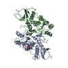
| Title | CDPK1 from Neospora caninum in complex with inhibitor RM-1-132 | ||||||
 Components Components | Calmodulin-like domain protein kinase isoenzyme gamma, related | ||||||
 Keywords Keywords | TRANSFERASE/TRANSFERASE INHIBITOR / serine/threonine protein kinase / transferase / calcium-binding / ATP-binding / TRANSFERASE-TRANSFERASE INHIBITOR complex | ||||||
| Function / homology |  Function and homology information Function and homology informationorganelle / non-specific serine/threonine protein kinase / protein serine/threonine kinase activity / calcium ion binding / ATP binding Similarity search - Function | ||||||
| Biological species |  Neospora caninum (eukaryote) Neospora caninum (eukaryote) | ||||||
| Method |  X-RAY DIFFRACTION / SYNCHROTRON / FOURIER SYNTHESIS / Resolution: 3 Å X-RAY DIFFRACTION / SYNCHROTRON / FOURIER SYNTHESIS / Resolution: 3 Å | ||||||
 Authors Authors | Merritt, E.A. | ||||||
 Citation Citation | Journal: Plos One / Year: 2014 Title: Neospora caninum Calcium-Dependent Protein Kinase 1 Is an Effective Drug Target for Neosporosis Therapy. Authors: Ojo, K.K. / Reid, M.C. / Kallur Siddaramaiah, L. / Muller, J. / Winzer, P. / Zhang, Z. / Keyloun, K.R. / Vidadala, R.S. / Merritt, E.A. / Hol, W.G. / Maly, D.J. / Fan, E. / Van Voorhis, W.C. / Hemphill, A. #1: Journal: Nat.Struct.Mol.Biol. / Year: 2010Title: Toxoplasma gondii calcium-dependent protein kinase 1 is a target for selective kinase inhibitors. Authors: Ojo, K.K. / Larson, E.T. / Keyloun, K.R. / Castaneda, L.J. / Derocher, A.E. / Inampudi, K.K. / Kim, J.E. / Arakaki, T.L. / Murphy, R.C. / Zhang, L. / Napuli, A.J. / Maly, D.J. / Verlinde, C. ...Authors: Ojo, K.K. / Larson, E.T. / Keyloun, K.R. / Castaneda, L.J. / Derocher, A.E. / Inampudi, K.K. / Kim, J.E. / Arakaki, T.L. / Murphy, R.C. / Zhang, L. / Napuli, A.J. / Maly, D.J. / Verlinde, C.L. / Buckner, F.S. / Parsons, M. / Hol, W.G. / Merritt, E.A. / Van Voorhis, W.C. | ||||||
| History |
|
- Structure visualization
| Structure viewer | Molecule: MolmilJmol/JSmol |
|---|
- Downloads & links
-Download
| PDBx/mmCIF format | 4mxa.cif.gz | 203.9 KB | Display | PDBx/mmCIF format |
|---|---|---|---|---|
| PDB format | pdb4mxa.ent.gz | 163.4 KB | Display | PDB format |
| PDBx/mmJSON format | 4mxa.json.gz | Tree view | PDBx/mmJSON format | |
| Others |  Other downloads Other downloads |
-Validation report
| Arichive directory | https://data.pdbj.org/pub/pdb/validation_reports/mx/4mxaftp://data.pdbj.org/pub/pdb/validation_reports/mx/4mxa | HTTPS FTP |
|---|
-Related structure data
| Related structure data | 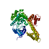 4m97SC 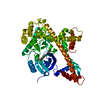 4mx9C S: Starting model for refinement C: citing same article ( |
|---|---|
| Similar structure data |


-Links
 PDBj
PDBj- Assembly
| Deposited unit | 
| ||||||||
|---|---|---|---|---|---|---|---|---|---|
| 1 |
| ||||||||
| Unit cell |
|
-Components
| #1: Protein | Mass: 55083.688 Da / Num. of mol.: 1 / Fragment: RESIDUES 22-506 Source method: isolated from a genetically manipulated source Source: (gene. exp.) Neospora caninum (eukaryote) / Strain: Liverpool / Gene: CDPK1, NCLIV_011980, XP_003880764 / Plasmid: AVA0421 / Production host:  References: UniProt: F0V9W9, Ca2+/calmodulin-dependent protein kinase |
|---|---|
| #2: Chemical | ChemComp-BK7 /   Mass: 402.492 Da / Num. of mol.: 1 / Source method: obtained synthetically / Formula: C23H26N6O Mass: 402.492 Da / Num. of mol.: 1 / Source method: obtained synthetically / Formula: C23H26N6O |
| #3: Water | ChemComp-HOH /  Mass: 18.015 Da / Num. of mol.: 25 / Source method: isolated from a natural source / Formula: H2O Mass: 18.015 Da / Num. of mol.: 25 / Source method: isolated from a natural source / Formula: H2O |
-Experimental details
-Experiment
| Experiment | Method: X-RAY DIFFRACTION / Number of used crystals: 1 |
|---|
- Sample preparation
| Crystal | Density Matthews: 2.06 Å3/Da / Density % sol: 40.2 % |
|---|---|
| Crystal grow | Temperature: 298 K / Method: vapor diffusion, sitting drop / pH: 7 Details: protein solution: 25 mM HEPES pH 7.0, 0.5 M NaCl, 5% glycerol, 5 mM DTT, 20 mM EGTA, 3mg/ml protein, 0.2 mM RM-1-132, 1% DMSO; crystallization buffer: 30% PEG 3350, 0.2 M ammonium citrate, 0. ...Details: protein solution: 25 mM HEPES pH 7.0, 0.5 M NaCl, 5% glycerol, 5 mM DTT, 20 mM EGTA, 3mg/ml protein, 0.2 mM RM-1-132, 1% DMSO; crystallization buffer: 30% PEG 3350, 0.2 M ammonium citrate, 0.1 M BisTris pH 5.3, 5 mM DTT, vapor diffusion, sitting drop, temperature 298K |
-Data collection
| Diffraction | Mean temperature: 100 K | |||||||||||||||
|---|---|---|---|---|---|---|---|---|---|---|---|---|---|---|---|---|
| Diffraction source | Source: SYNCHROTRON / Site: SSRL  / Beamline: BL12-2 / Wavelength: 0.97939 Å / Beamline: BL12-2 / Wavelength: 0.97939 Å | |||||||||||||||
| Detector | Type: DECTRIS PILATUS 6M / Detector: PIXEL / Date: Jun 6, 2013 | |||||||||||||||
| Radiation | Monochromator: Side scattering bent cube-root I-beam single crystal; asymmetric cut 4.965 degs. Protocol: SINGLE WAVELENGTH / Monochromatic (M) / Laue (L): M / Scattering type: x-ray | |||||||||||||||
| Radiation wavelength | Wavelength: 0.97939 Å / Relative weight: 1 | |||||||||||||||
| Reflection | Resolution: 3→47.59 Å / Num. obs: 8027 / % possible obs: 89.1 % / Redundancy: 2.8 % / Rmerge(I) obs: 0.376 / Net I/σ(I): 3.6 | |||||||||||||||
| Reflection shell | Rmerge(I) obs: 0.018 / Diffraction-ID: 1 / Redundancy: 2.7 %
|
- Processing
| Software |
| |||||||||||||||||||||||||||||||||||||||||||||||||||||||||||||||||||||||||||||||||||||||||||||||||||||||||||||||||||||||||||||||||||||||||||||||||||||||||||||||||||||||||||||||
|---|---|---|---|---|---|---|---|---|---|---|---|---|---|---|---|---|---|---|---|---|---|---|---|---|---|---|---|---|---|---|---|---|---|---|---|---|---|---|---|---|---|---|---|---|---|---|---|---|---|---|---|---|---|---|---|---|---|---|---|---|---|---|---|---|---|---|---|---|---|---|---|---|---|---|---|---|---|---|---|---|---|---|---|---|---|---|---|---|---|---|---|---|---|---|---|---|---|---|---|---|---|---|---|---|---|---|---|---|---|---|---|---|---|---|---|---|---|---|---|---|---|---|---|---|---|---|---|---|---|---|---|---|---|---|---|---|---|---|---|---|---|---|---|---|---|---|---|---|---|---|---|---|---|---|---|---|---|---|---|---|---|---|---|---|---|---|---|---|---|---|---|---|---|---|---|---|
| Refinement | Method to determine structure: FOURIER SYNTHESIS Starting model: PDB ENTRY 4M97 Resolution: 3→47.63 Å / Cor.coef. Fo:Fc: 0.853 / Cor.coef. Fo:Fc free: 0.807 / WRfactor Rfree: 0.2815 / WRfactor Rwork: 0.2577 / Occupancy max: 1 / Occupancy min: 0.3 / FOM work R set: 0.7868 / SU B: 60.368 / SU ML: 0.555 / SU R Cruickshank DPI: 0.726 / SU Rfree: 0.6883 / Cross valid method: THROUGHOUT / σ(F): 0 / ESU R Free: 0.688 / Stereochemistry target values: MAXIMUM LIKELIHOOD Details: HYDROGENS HAVE BEEN ADDED IN THE RIDING POSITIONS U VALUES : WITH TLS ADDED
| |||||||||||||||||||||||||||||||||||||||||||||||||||||||||||||||||||||||||||||||||||||||||||||||||||||||||||||||||||||||||||||||||||||||||||||||||||||||||||||||||||||||||||||||
| Solvent computation | Ion probe radii: 0.8 Å / Shrinkage radii: 0.8 Å / VDW probe radii: 1.2 Å / Solvent model: MASK | |||||||||||||||||||||||||||||||||||||||||||||||||||||||||||||||||||||||||||||||||||||||||||||||||||||||||||||||||||||||||||||||||||||||||||||||||||||||||||||||||||||||||||||||
| Displacement parameters | Biso max: 202.59 Å2 / Biso mean: 73.6112 Å2 / Biso min: 2 Å2
| |||||||||||||||||||||||||||||||||||||||||||||||||||||||||||||||||||||||||||||||||||||||||||||||||||||||||||||||||||||||||||||||||||||||||||||||||||||||||||||||||||||||||||||||
| Refinement step | Cycle: LAST / Resolution: 3→47.63 Å
| |||||||||||||||||||||||||||||||||||||||||||||||||||||||||||||||||||||||||||||||||||||||||||||||||||||||||||||||||||||||||||||||||||||||||||||||||||||||||||||||||||||||||||||||
| Refine LS restraints |
| |||||||||||||||||||||||||||||||||||||||||||||||||||||||||||||||||||||||||||||||||||||||||||||||||||||||||||||||||||||||||||||||||||||||||||||||||||||||||||||||||||||||||||||||
| LS refinement shell | Resolution: 3→3.078 Å / Total num. of bins used: 20
| |||||||||||||||||||||||||||||||||||||||||||||||||||||||||||||||||||||||||||||||||||||||||||||||||||||||||||||||||||||||||||||||||||||||||||||||||||||||||||||||||||||||||||||||
| Refinement TLS params. | Method: refined / Refine-ID: X-RAY DIFFRACTION
| |||||||||||||||||||||||||||||||||||||||||||||||||||||||||||||||||||||||||||||||||||||||||||||||||||||||||||||||||||||||||||||||||||||||||||||||||||||||||||||||||||||||||||||||
| Refinement TLS group |
|
