Mass: 18.015 Da / Num. of mol.: 39 / Source method: isolated from a natural source / Formula: H2O
Has protein modification
Y
-
Experimental details
-
Experiment
Experiment
Method: X-RAY DIFFRACTION / Number of used crystals: 1
-
Sample preparation
Crystal
Density Matthews: 2.7 Å3/Da / Density % sol: 54.49 % / Description: NONE
Crystal grow
Temperature: 293 K / Method: vapor diffusion, hanging drop / pH: 7 Details: CRB2-BRCT2 (3.5MG/ML IN 0.05M TRIS-HCL PH7.0, 0.6M NACL, 5MM DTT) WAS MIXED WITH AN EQUAL VOLUME OF PEPTIDE (0.05M TRIS-HCL PH7.0, 0.1M NACL, 5MM DTT), TO GIVE A 1:30 MOLAR RATIO OF PROTEIN: ...Details: CRB2-BRCT2 (3.5MG/ML IN 0.05M TRIS-HCL PH7.0, 0.6M NACL, 5MM DTT) WAS MIXED WITH AN EQUAL VOLUME OF PEPTIDE (0.05M TRIS-HCL PH7.0, 0.1M NACL, 5MM DTT), TO GIVE A 1:30 MOLAR RATIO OF PROTEIN:PEPTIDE. CRB2-BRCT2/PEPTIDE (7MG/ML IN 0.05M TRIS-HCL PH7.0, 1M NACL, 5MM DTT) WERE CRYSTALLISED BY HANGING DROP VAPOUR DIFFUSION AT 20C. PLATECLUSTERS GREW FROM 2UL PROTEIN, 2UL WELL SOLUTION (0.1M NA-CACODYLATE PH6.5, 7.0% PEG8000 AND 0.2M CA-ACETATE) AND 0.5UL 0.1M PR(III) ACETATE.
Protocol: SINGLE WAVELENGTH / Monochromatic (M) / Laue (L): M / Scattering type: x-ray
Radiation wavelength
Wavelength: 1.06 Å / Relative weight: 1
Reflection
Resolution: 3.1→70 Å / Num. obs: 11193 / % possible obs: 99.8 % / Observed criterion σ(I): 0 / Redundancy: 3.8 % / Biso Wilson estimate: 53.11 Å2 / Rmerge(I) obs: 0.09 / Net I/σ(I): 11.5
Reflection shell
Resolution: 3.1→3.27 Å / Redundancy: 4 % / Rmerge(I) obs: 0.35 / Mean I/σ(I) obs: 3.7 / % possible all: 100
-
Processing
Software
Name
Version
Classification
PHENIX
(PHENIX.REFINE)
refinement
MOSFLM
datareduction
SCALA
datascaling
PHASER
phasing
Refinement
Method to determine structure: MOLECULAR REPLACEMENT Starting model: IN HOUSE STRUCTURE Resolution: 3.1→65 Å / SU ML: 0.41 / Phase error: 22.54 / Stereochemistry target values: ML
Rfactor
Num. reflection
% reflection
Rfree
0.2444
968
4.8 %
Rwork
0.1952
-
-
obs
0.1976
20371
98.9 %
Solvent computation
Shrinkage radii: 0.9 Å / VDW probe radii: 1.11 Å / Solvent model: FLAT BULK SOLVENT MODEL / Bsol: 56.99 Å2 / ksol: 0.3 e/Å3
Displacement parameters
Biso mean: 57.77 Å2
Baniso -1
Baniso -2
Baniso -3
1-
-1.3932 Å2
0 Å2
0 Å2
2-
-
4.1243 Å2
-0 Å2
3-
-
-
-2.731 Å2
Refinement step
Cycle: LAST / Resolution: 3.1→65 Å
Protein
Nucleic acid
Ligand
Solvent
Total
Num. atoms
3517
0
4
39
3560
Refine LS restraints
Refine-ID
Type
Dev ideal
Number
X-RAY DIFFRACTION
f_bond_d
0.007
3598
X-RAY DIFFRACTION
f_angle_d
1.047
4903
X-RAY DIFFRACTION
f_dihedral_angle_d
16.849
1257
X-RAY DIFFRACTION
f_chiral_restr
0.07
567
X-RAY DIFFRACTION
f_plane_restr
0.005
636
Refine LS restraints NCS
Ens-ID
Dom-ID
Auth asym-ID
Number
Refine-ID
Type
Rms dev position (Å)
1
1
A
1653
X-RAY DIFFRACTION
POSITIONAL
1
2
B
1653
X-RAY DIFFRACTION
POSITIONAL
0.039
LS refinement shell
Resolution (Å)
Rfactor Rfree
Num. reflection Rfree
Rfactor Rwork
Num. reflection Rwork
Refine-ID
% reflection obs (%)
3.1-3.2634
0.3044
152
0.2475
2741
X-RAY DIFFRACTION
100
3.2634-3.4679
0.2963
145
0.2204
2819
X-RAY DIFFRACTION
100
3.4679-3.7356
0.272
135
0.2049
2766
X-RAY DIFFRACTION
99
3.7356-4.1115
0.2805
129
0.168
2808
X-RAY DIFFRACTION
99
4.1115-4.7063
0.1574
140
0.1441
2770
X-RAY DIFFRACTION
99
4.7063-5.9288
0.1944
128
0.1721
2745
X-RAY DIFFRACTION
98
5.9288-65.0335
0.244
139
0.2189
2754
X-RAY DIFFRACTION
98
Refinement TLS params.
Method: refined / Refine-ID: X-RAY DIFFRACTION
ID
L11 (°2)
L12 (°2)
L13 (°2)
L22 (°2)
L23 (°2)
L33 (°2)
S11 (Å °)
S12 (Å °)
S13 (Å °)
S21 (Å °)
S22 (Å °)
S23 (Å °)
S31 (Å °)
S32 (Å °)
S33 (Å °)
T11 (Å2)
T12 (Å2)
T13 (Å2)
T22 (Å2)
T23 (Å2)
T33 (Å2)
Origin x (Å)
Origin y (Å)
Origin z (Å)
1
0.9561
-0.0849
-0.0212
0.8951
0.3206
2.2293
0.0732
-0.1587
-0.1925
0.0005
0.0014
-0.0421
0.4034
-0.0434
-0.0001
0.1436
0.0157
-0.0299
0.1302
-0.0293
0.2257
-17.0659
19.2577
-4.9341
2
0.7816
0.7286
0.5338
1.1622
0.0541
0.7944
-0.1495
0.42
-0.0142
-0.4505
0.0768
-0.0088
-0.1027
0.352
-0.0008
0.343
-0.0067
0.0857
0.2918
-0.0198
0.1527
-1.1531
38.8413
-21.0865
Refinement TLS group
ID
Refine-ID
Refine TLS-ID
Selection details
1
X-RAY DIFFRACTION
1
CHAINA
2
X-RAY DIFFRACTION
2
CHAINB
+
About Yorodumi
-
News
-
Feb 9, 2022. New format data for meta-information of EMDB entries
New format data for meta-information of EMDB entries
Version 3 of the EMDB header file is now the official format.
The previous official version 1.9 will be removed from the archive.
In the structure databanks used in Yorodumi, some data are registered as the other names, "COVID-19 virus" and "2019-nCoV". Here are the details of the virus and the list of structure data.
Jan 31, 2019. EMDB accession codes are about to change! (news from PDBe EMDB page)
EMDB accession codes are about to change! (news from PDBe EMDB page)
The allocation of 4 digits for EMDB accession codes will soon come to an end. Whilst these codes will remain in use, new EMDB accession codes will include an additional digit and will expand incrementally as the available range of codes is exhausted. The current 4-digit format prefixed with “EMD-” (i.e. EMD-XXXX) will advance to a 5-digit format (i.e. EMD-XXXXX), and so on. It is currently estimated that the 4-digit codes will be depleted around Spring 2019, at which point the 5-digit format will come into force.
The EM Navigator/Yorodumi systems omit the EMD- prefix.
Related info.:Q: What is EMD? / ID/Accession-code notation in Yorodumi/EM Navigator
Yorodumi is a browser for structure data from EMDB, PDB, SASBDB, etc.
This page is also the successor to EM Navigator detail page, and also detail information page/front-end page for Omokage search.
The word "yorodu" (or yorozu) is an old Japanese word meaning "ten thousand". "mi" (miru) is to see.
Related info.:EMDB / PDB / SASBDB / Comparison of 3 databanks / Yorodumi Search / Aug 31, 2016. New EM Navigator & Yorodumi / Yorodumi Papers / Jmol/JSmol / Function and homology information / Changes in new EM Navigator and Yorodumi
 Movie
Movie Controller


 Open data
 Basic information
 Components
 Keywords
 Function and homology information
 HOMO SAPIENS (human)
 X-RAY DIFFRACTION /
  Authors
 Citation
 Structure visualization
 Downloads & links
 Other downloads


 PDBj
PDBj

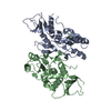
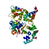
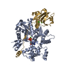
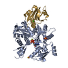
 Assembly
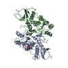

 Mass: 140.908 Da / Num. of mol.: 4 / Source method: obtained synthetically / Formula: Pr
 Mass: 18.015 Da / Num. of mol.: 39 / Source method: isolated from a natural source / Formula: H2O
 Sample preparation
 / Beamline: I03 / Wavelength: 1.06
  Processing