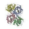
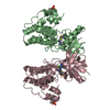
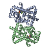
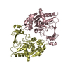
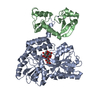
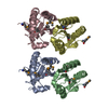
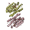
- PDB-4mud: Crystal structure of a ring oxydation complex/ phenylacetic acid ... -
+
Open data
ID or keywords:
Loading...
-
Basic information
Entry
Database: PDB / ID: 4mud
Title
Crystal structure of a ring oxydation complex/ phenylacetic acid degradation-like protein (SSO1313) from Sulfolobus solfataricus P2 at 2.43 A resolution
Components
Ring oxydation complex/ phenylacetic acid degradation related protein
Keywords
OXIDOREDUCTASE / Phenylacetic acid catabolic protein / PF05138 family / Structural Genomics / Joint Center for Structural Genomics / JCSG / Protein Structure Initiative / PSI-BIOLOGY
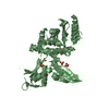
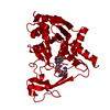
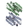
A: Ring oxydation complex/ phenylacetic acid degradation related protein B: Ring oxydation complex/ phenylacetic acid degradation related protein C: Ring oxydation complex/ phenylacetic acid degradation related protein D: Ring oxydation complex/ phenylacetic acid degradation related protein hetero molecules
C: Ring oxydation complex/ phenylacetic acid degradation related protein D: Ring oxydation complex/ phenylacetic acid degradation related protein hetero molecules
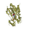
Mass: 18.015 Da / Num. of mol.: 170 / Source method: isolated from a natural source / Formula: H2O
Has protein modification
Y
Sequence details
THE CONSTRUCT WAS EXPRESSED WITH A PURIFICATION TAG MGSDKIHHHHHHENLYFQG. THE TAG WAS REMOVED WITH ...THE CONSTRUCT WAS EXPRESSED WITH A PURIFICATION TAG MGSDKIHHHHHHENLYFQG. THE TAG WAS REMOVED WITH TEV PROTEASE LEAVING ONLY A GLYCINE (0) FOLLOWED BY RESIDUES 1-234 OF THE TARGET SEQUENCE.
-
Experimental details
-
Experiment
Experiment
Method: X-RAY DIFFRACTION / Number of used crystals: 1
-
Sample preparation
Crystal
Density Matthews: 2.19 Å3/Da / Density % sol: 43.73 %
Crystal grow
Temperature: 277 K / Method: vapor diffusion, sitting drop / pH: 7.2 Details: 20.00% polyethylene glycol 3350, 0.200M sodium formate, No Buffer pH 7.2, Additive: 0.003 M phenylacetate, NANODROP', VAPOR DIFFUSION, SITTING DROP, temperature 277K
Type: DECTRIS PILATUS 6M / Detector: PIXEL / Date: May 22, 2013 Details: Flat mirror (vertical focusing); single crystal Si(111) bent monochromator (horizontal focusing)
Radiation
Monochromator: single crystal Si(111) bent / Protocol: MAD / Monochromatic (M) / Laue (L): M / Scattering type: x-ray
Radiation wavelength
ID
Wavelength (Å)
Relative weight
1
0.91837
1
2
0.97941
1
3
0.97855
1
Reflection
Resolution: 2.43→29.229 Å / Num. obs: 31723 / % possible obs: 86.7 % / Observed criterion σ(I): -3 / Biso Wilson estimate: 45.633 Å2 / Rmerge(I) obs: 0.08 / Net I/σ(I): 5
Reflection shell
Diffraction-ID: 1
Resolution (Å)
Highest resolution (Å)
Rmerge(I) obs
Mean I/σ(I) obs
Num. measured obs
Num. unique obs
% possible all
2.43-2.52
0.224
1.9
7327
6445
87.7
2.52-2.62
0.184
2.2
6881
6096
87.8
2.62-2.74
0.164
2.6
6992
6192
87.6
2.74-2.88
0.129
3.4
6703
5928
86.2
2.88-3.06
0.116
4.2
6289
5589
78.3
3.06-3.3
0.094
5.2
7236
6452
90.7
3.3-3.62
0.09
6.5
6804
6128
90
3.62-4.15
0.074
7.6
6938
6282
87.1
4.15-5.21
0.052
8.1
6502
5891
83.9
5.21
0.057
8.3
6965
6354
88.2
-
Phasing
Phasing
Method: MAD
-
Processing
Software
Name
Version
Classification
NB
MolProbity
3beta29
modelbuilding
PDB_EXTRACT
3.1
dataextraction
SOLVE
phasing
XSCALE
July4, 2012
datascaling
BUSTER-TNT
2.10.0
refinement
XDS
datareduction
BUSTER
2.10.0
refinement
Refinement
Method to determine structure: MAD / Resolution: 2.43→29.229 Å / Cor.coef. Fo:Fc: 0.939 / Cor.coef. Fo:Fc free: 0.922 / Occupancy max: 1 / Occupancy min: 0.33 / Cross valid method: THROUGHOUT / σ(F): 0 Details: 1. A MET-INHIBITION PROTOCOL WAS USED FOR SELENOMETHIONINE INCORPORATION DURING PROTEIN EXPRESSION. THE OCCUPANCY OF THE SE ATOMS IN THE MSE RESIDUES WAS REDUCED TO 0.75 FOR THE REDUCED ...Details: 1. A MET-INHIBITION PROTOCOL WAS USED FOR SELENOMETHIONINE INCORPORATION DURING PROTEIN EXPRESSION. THE OCCUPANCY OF THE SE ATOMS IN THE MSE RESIDUES WAS REDUCED TO 0.75 FOR THE REDUCED SCATTERING POWER DUE TO PARTIAL S-MET INCORPORATION. 2. ATOM RECORD CONTAINS SUM OF TLS AND RESIDUAL B FACTORS. ANISOU RECORD CONTAINS SUM OF TLS AND RESIDUAL U FACTORS. 3. THE MAD PHASES WERE USED AS RESTRAINTS DURING REFINEMENT. 4. NCS RESTRAINTS WERE APPLIED USING BUSTER'S LSSR RESTRAINT REPRESENTATION (-AUTONCS). 5.1,2-ETHANEDIOL (EDO) USED AS A CRYOPROTECTANT WERE MODELED INTO THE STRUCTURE.
In the structure databanks used in Yorodumi, some data are registered as the other names, "COVID-19 virus" and "2019-nCoV". Here are the details of the virus and the list of structure data.
Jan 31, 2019. EMDB accession codes are about to change! (news from PDBe EMDB page)
EMDB accession codes are about to change! (news from PDBe EMDB page)
The allocation of 4 digits for EMDB accession codes will soon come to an end. Whilst these codes will remain in use, new EMDB accession codes will include an additional digit and will expand incrementally as the available range of codes is exhausted. The current 4-digit format prefixed with “EMD-” (i.e. EMD-XXXX) will advance to a 5-digit format (i.e. EMD-XXXXX), and so on. It is currently estimated that the 4-digit codes will be depleted around Spring 2019, at which point the 5-digit format will come into force.
The EM Navigator/Yorodumi systems omit the EMD- prefix.
Related info.:Q: What is EMD? / ID/Accession-code notation in Yorodumi/EM Navigator
Yorodumi is a browser for structure data from EMDB, PDB, SASBDB, etc.
This page is also the successor to EM Navigator detail page, and also detail information page/front-end page for Omokage search.
The word "yorodu" (or yorozu) is an old Japanese word meaning "ten thousand". "mi" (miru) is to see.
Related info.:EMDB / PDB / SASBDB / Comparison of 3 databanks / Yorodumi Search / Aug 31, 2016. New EM Navigator & Yorodumi / Yorodumi Papers / Jmol/JSmol / Function and homology information / Changes in new EM Navigator and Yorodumi
 Movie
Movie Controller

 Yorodumi
 Open data
 Basic information
 Components
 Keywords
 Function and homology information
 Sulfolobus solfataricus (archaea)
 X-RAY DIFFRACTION /
  Authors
 Citation
 Structure visualization
 Downloads & links
 Other downloads
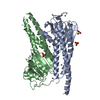
 PDBj
PDBj


 Assembly
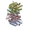


 Mass: 62.068 Da / Num. of mol.: 8 / Source method: obtained synthetically / Formula: C2H6O2
 Mass: 18.015 Da / Num. of mol.: 170 / Source method: isolated from a natural source / Formula: H2O
 Sample preparation
 / Beamline: BL11-1 / Wavelength: 0.91837,0.97941,0.97855
 Processing