 Movie
Movie Controller

[English] 日本語
 Yorodumi
- PDB-4mrt: Structure of the Phosphopantetheine Transferase Sfp in Complex wi... -
+ Open data

- Basic information
| Entry | Database: PDB / ID: 4mrt | ||||||
|---|---|---|---|---|---|---|---|
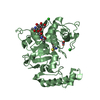
| Title | Structure of the Phosphopantetheine Transferase Sfp in Complex with Coenzyme A and a Peptidyl Carrier Protein | ||||||
 Components Components |
| ||||||
 Keywords Keywords | TRANSPORT PROTEIN/TRANSFERASE / PCP: Aminoacyl/peptidyl carrier / Sfp: Phosphopantetheine transferase / Sfp: Coenzyme A binding / TRANSPORT PROTEIN-TRANSFERASE complex | ||||||
| Function / homology |  Function and homology information Function and homology informationpositive regulation of single-species biofilm formation / holo-[acyl-carrier-protein] synthase / : / holo-[acyl-carrier-protein] synthase activity / amino acid activation for nonribosomal peptide biosynthetic process / secondary metabolite biosynthetic process / lipid biosynthetic process / ligase activity / phosphopantetheine binding / antibiotic biosynthetic process ...positive regulation of single-species biofilm formation / holo-[acyl-carrier-protein] synthase / : / holo-[acyl-carrier-protein] synthase activity / amino acid activation for nonribosomal peptide biosynthetic process / secondary metabolite biosynthetic process / lipid biosynthetic process / ligase activity / phosphopantetheine binding / antibiotic biosynthetic process / fatty acid biosynthetic process / magnesium ion binding / cytosol Similarity search - Function | ||||||
| Biological species |  Brevibacillus parabrevis (bacteria) Brevibacillus parabrevis (bacteria) | ||||||
| Method |  X-RAY DIFFRACTION / SYNCHROTRON / MOLECULAR REPLACEMENT / molecular replacement / Resolution: 2 Å X-RAY DIFFRACTION / SYNCHROTRON / MOLECULAR REPLACEMENT / molecular replacement / Resolution: 2 Å | ||||||
 Authors Authors | Tufar, P. / Rahighi, S. / Kraas, F.I. / Kirchner, D.K. / Loehr, F. / Henrich, E. / Koepke, J. / Dikic, I. / Guentert, P. / Marahiel, M.A. / Doetsch, V. | ||||||
 Citation Citation | Journal: Chem.Biol. / Year: 2014 Title: Crystal Structure of a PCP/Sfp Complex Reveals the Structural Basis for Carrier Protein Posttranslational Modification. Authors: Tufar, P. / Rahighi, S. / Kraas, F.I. / Kirchner, D.K. / Lohr, F. / Henrich, E. / Kopke, J. / Dikic, I. / Guntert, P. / Marahiel, M.A. / Dotsch, V. | ||||||
| History |
|
- Structure visualization
| Structure viewer | Molecule: MolmilJmol/JSmol |
|---|
- Downloads & links
-Download
| PDBx/mmCIF format | 4mrt.cif.gz | 82.6 KB | Display | PDBx/mmCIF format |
|---|---|---|---|---|
| PDB format | pdb4mrt.ent.gz | 60.4 KB | Display | PDB format |
| PDBx/mmJSON format | 4mrt.json.gz | Tree view | PDBx/mmJSON format | |
| Others |  Other downloads Other downloads |
-Validation report
| Arichive directory | https://data.pdbj.org/pub/pdb/validation_reports/mr/4mrtftp://data.pdbj.org/pub/pdb/validation_reports/mr/4mrt | HTTPS FTP |
|---|
-Related structure data


-Links
 PDBj
PDBj


- Assembly
| Deposited unit | 
| ||||||||
|---|---|---|---|---|---|---|---|---|---|
| 1 |
| ||||||||
| Unit cell |
|
-Components
-Protein , 2 types, 2 molecules CA
| #1: Protein | Mass: 9972.424 Da / Num. of mol.: 1 Fragment: Peptidyl carrier protein domain(UNP Residues 3038-3113) Mutation: S3075A Source method: isolated from a genetically manipulated source Details: Cytosol / Source: (gene. exp.) Brevibacillus parabrevis (bacteria) / Strain: ATCC 8185 / Gene: tycC / Plasmid: pQE70 / Production host: |
|---|---|
| #2: Protein | Mass: 27245.822 Da / Num. of mol.: 1 Source method: isolated from a genetically manipulated source Details: Cytosol Source: (gene. exp.) Strain: 168 / Gene: BSU03570, lpa-8, sfp / Plasmid: pQE60 / Production host: References: UniProt: P39135, Transferases; Transferring phosphorus-containing groups; Transferases for other substituted phosphate groups |
-Non-polymers , 5 types, 177 molecules 


| #3: Chemical |  Mass: 24.305 Da / Num. of mol.: 2 / Source method: obtained synthetically / Formula: Mg Mass: 24.305 Da / Num. of mol.: 2 / Source method: obtained synthetically / Formula: Mg#4: Chemical | ChemComp-COA / |  Mass: 767.534 Da / Num. of mol.: 1 / Source method: obtained synthetically / Formula: C21H36N7O16P3S Mass: 767.534 Da / Num. of mol.: 1 / Source method: obtained synthetically / Formula: C21H36N7O16P3S#5: Chemical | ChemComp-GOL / |  Mass: 92.094 Da / Num. of mol.: 1 / Source method: obtained synthetically / Formula: C3H8O3 Mass: 92.094 Da / Num. of mol.: 1 / Source method: obtained synthetically / Formula: C3H8O3#6: Chemical | ChemComp-SO4 / |  Mass: 96.063 Da / Num. of mol.: 1 / Source method: obtained synthetically / Formula: SO4 Mass: 96.063 Da / Num. of mol.: 1 / Source method: obtained synthetically / Formula: SO4#7: Water | ChemComp-HOH / | Mass: 18.015 Da / Num. of mol.: 172 / Source method: isolated from a natural source / Formula: H2O |
|---|
-Experimental details
-Experiment
| Experiment | Method: X-RAY DIFFRACTION / Number of used crystals: 1 |
|---|
- Sample preparation
| Crystal | Density Matthews: 2.3 Å3/Da / Density % sol: 46.57 % |
|---|---|
| Crystal grow | Temperature: 293 K / Method: vapor diffusion, hanging drop / pH: 6.5 Details: 0.16 M magnesium acetate, 0.08 M sodium cacodylate, 16% (w/v) PEG 8000, 20% (v/v) glycerol, pH 6.5, VAPOR DIFFUSION, HANGING DROP, temperature 293K |
-Data collection
| Diffraction | Mean temperature: 100 K |
|---|---|
| Diffraction source | Source: SYNCHROTRON / Site: SLS  / Beamline: X06DA / Wavelength: 1 Å / Beamline: X06DA / Wavelength: 1 Å |
| Detector | Type: PSI PILATUS 6M / Detector: PIXEL / Date: Dec 8, 2012 Details: The optical elements consist of a vertically collimating mirror (M1, focus at infinity), followed by a Bartels Monochromator with dual channel cut crystals (DCCM) in (+--+) geometry, and a ...Details: The optical elements consist of a vertically collimating mirror (M1, focus at infinity), followed by a Bartels Monochromator with dual channel cut crystals (DCCM) in (+--+) geometry, and a toroidal mirror (M2) to vertically and horizontally focus the beam at the sample position (with 2:1 horizontal demagnification). |
| Radiation | Monochromator: Bartels Monochromator with dual channel cut crystals (DCCM) in (+--+) geometry Protocol: SINGLE WAVELENGTH / Monochromatic (M) / Laue (L): M / Scattering type: x-ray |
| Radiation wavelength | Wavelength: 1 Å / Relative weight: 1 |
| Reflection | Resolution: 2→37.91 Å / Num. all: 21610 / Num. obs: 21594 / % possible obs: 99.45 % / Observed criterion σ(F): 2 / Observed criterion σ(I): 2 / Redundancy: 6.2 % / Net I/σ(I): 6.7 |
-Phasing
| Phasing | Method: molecular replacement |
|---|
- Processing
| Software |
| ||||||||||||||||||||||||||||||||||||||||||||||||||||||||||||
|---|---|---|---|---|---|---|---|---|---|---|---|---|---|---|---|---|---|---|---|---|---|---|---|---|---|---|---|---|---|---|---|---|---|---|---|---|---|---|---|---|---|---|---|---|---|---|---|---|---|---|---|---|---|---|---|---|---|---|---|---|---|
| Refinement | Method to determine structure: MOLECULAR REPLACEMENT / Resolution: 2→37.91 Å / Cor.coef. Fo:Fc: 0.954 / Cor.coef. Fo:Fc free: 0.934 / WRfactor Rfree: 0.2531 / WRfactor Rwork: 0.2036 / Occupancy max: 1 / Occupancy min: 0.5 / FOM work R set: 0.8521 / SU B: 4.467 / SU ML: 0.124 / SU R Cruickshank DPI: 0.1998 / SU Rfree: 0.1709 / Cross valid method: THROUGHOUT / σ(F): 0 / ESU R: 0.2 / ESU R Free: 0.171 / Stereochemistry target values: MAXIMUM LIKELIHOOD Details: HYDROGENS HAVE BEEN ADDED IN THE RIDING POSITIONS U VALUES : REFINED INDIVIDUALLY
| ||||||||||||||||||||||||||||||||||||||||||||||||||||||||||||
| Solvent computation | Ion probe radii: 0.8 Å / Shrinkage radii: 0.8 Å / VDW probe radii: 1.2 Å / Solvent model: MASK | ||||||||||||||||||||||||||||||||||||||||||||||||||||||||||||
| Displacement parameters | Biso max: 70.62 Å2 / Biso mean: 29.0043 Å2 / Biso min: 10.07 Å2
| ||||||||||||||||||||||||||||||||||||||||||||||||||||||||||||
| Refinement step | Cycle: LAST / Resolution: 2→37.91 Å
| ||||||||||||||||||||||||||||||||||||||||||||||||||||||||||||
| Refine LS restraints |
| ||||||||||||||||||||||||||||||||||||||||||||||||||||||||||||
| LS refinement shell | Resolution: 2→2.052 Å / Total num. of bins used: 20
|
