 Movie
Movie Controller

[English] 日本語
 Yorodumi
- PDB-4mqd: Crystal structure of ComJ, inhibitor of the DNA degrading activit... -
+ Open data

- Basic information
| Entry | Database: PDB / ID: 4mqd | ||||||
|---|---|---|---|---|---|---|---|
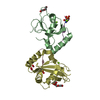
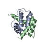
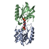
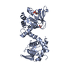
| Title | Crystal structure of ComJ, inhibitor of the DNA degrading activity of NucA, from Bacillus subtilis | ||||||
 Components Components | DNA-entry nuclease inhibitor | ||||||
 Keywords Keywords | Hydrolase Inhibitor / inhibitor of NucA / Competence protein J / Midwest center for structural genomics / MCSG / PSI-BIOLOGY | ||||||
| Function / homology |  Function and homology information Function and homology information | ||||||
| Biological species |  | ||||||
| Method |  X-RAY DIFFRACTION / SYNCHROTRON / SAD / Resolution: 2.16 Å X-RAY DIFFRACTION / SYNCHROTRON / SAD / Resolution: 2.16 Å | ||||||
 Authors Authors | Chang, C. / Mack, J. / Clancy, S. / Joachimiak, A. / Midwest Center for Structural Genomics (MCSG) | ||||||
 Citation Citation | Journal: To be Published Title: Crystal structure of ComJ, inhibitor of the DNA degrading activity of NucA, from Bacillus subtilis Authors: Chang, C. / Mack, J. / Clancy, S. / Joachimiak, A. | ||||||
| History |
|
- Structure visualization
| Structure viewer | Molecule: MolmilJmol/JSmol |
|---|
- Downloads & links
-Download
| PDBx/mmCIF format | 4mqd.cif.gz | 233.6 KB | Display | PDBx/mmCIF format |
|---|---|---|---|---|
| PDB format | pdb4mqd.ent.gz | 191.1 KB | Display | PDB format |
| PDBx/mmJSON format | 4mqd.json.gz | Tree view | PDBx/mmJSON format | |
| Others |  Other downloads Other downloads |
-Validation report
| Arichive directory | https://data.pdbj.org/pub/pdb/validation_reports/mq/4mqdftp://data.pdbj.org/pub/pdb/validation_reports/mq/4mqd | HTTPS FTP |
|---|
-Related structure data
| Similar structure data | |
|---|---|
| Other databases |


-Links
 PDBj
PDBj- Assembly
| Deposited unit | 
| ||||||||
|---|---|---|---|---|---|---|---|---|---|
| 1 | 
| ||||||||
| 2 | 
| ||||||||
| 3 | 
| ||||||||
| 4 | 
| ||||||||
| Unit cell |
| ||||||||
| Details | A and B, C and D from PISA |
-Components
| #1: Protein | Mass: 15472.742 Da / Num. of mol.: 4 Source method: isolated from a genetically manipulated source Source: (gene. exp.) Strain: 168 / Gene: nin, comJ, BSU03420 / Plasmid: pMCSG73 / Production host: #2: Water | ChemComp-HOH / |  Mass: 18.015 Da / Num. of mol.: 269 / Source method: isolated from a natural source / Formula: H2O Mass: 18.015 Da / Num. of mol.: 269 / Source method: isolated from a natural source / Formula: H2OHas protein modification | Y | |
|---|
-Experimental details
-Experiment
| Experiment | Method: X-RAY DIFFRACTION / Number of used crystals: 1 |
|---|
- Sample preparation
| Crystal | Density Matthews: 2.73 Å3/Da / Density % sol: 55 % |
|---|---|
| Crystal grow | Temperature: 297 K / Method: vapor diffusion, sitting drop / pH: 4.5 Details: 0.1M sodium acetate pH 4.5, 20% PEG 3000, VAPOR DIFFUSION, SITTING DROP, temperature 297K |
-Data collection
| Diffraction | Mean temperature: 100 K |
|---|---|
| Diffraction source | Source: SYNCHROTRON / Site: APS  / Beamline: 19-ID / Wavelength: 0.97929 Å / Beamline: 19-ID / Wavelength: 0.97929 Å |
| Detector | Type: ADSC QUANTUM 315r / Detector: CCD / Date: Feb 7, 2013 |
| Radiation | Monochromator: Si(111) double crystal / Protocol: SINGLE WAVELENGTH / Monochromatic (M) / Laue (L): M / Scattering type: x-ray |
| Radiation wavelength | Wavelength: 0.97929 Å / Relative weight: 1 |
| Reflection | Resolution: 2.15→50 Å / Num. all: 35143 / Num. obs: 35026 / % possible obs: 99.7 % / Observed criterion σ(I): -3 / Redundancy: 4.6 % / Rmerge(I) obs: 0.111 / Net I/σ(I): 12.3 |
| Reflection shell | Resolution: 2.15→2.19 Å / Redundancy: 4.1 % / Rmerge(I) obs: 0.974 / Mean I/σ(I) obs: 1.54 / Num. unique all: 1786 / % possible all: 99.9 |
- Processing
| Software |
| |||||||||||||||||||||||||||||||||||||||||||||||||||||||||||||||||||||||||||
|---|---|---|---|---|---|---|---|---|---|---|---|---|---|---|---|---|---|---|---|---|---|---|---|---|---|---|---|---|---|---|---|---|---|---|---|---|---|---|---|---|---|---|---|---|---|---|---|---|---|---|---|---|---|---|---|---|---|---|---|---|---|---|---|---|---|---|---|---|---|---|---|---|---|---|---|---|
| Refinement | Method to determine structure: SAD / Resolution: 2.16→41.22 Å / Cor.coef. Fo:Fc: 0.953 / Cor.coef. Fo:Fc free: 0.915 / Occupancy max: 1 / Occupancy min: 0.5 / SU B: 13.221 / SU ML: 0.15 / Cross valid method: THROUGHOUT / σ(F): 0 / ESU R Free: 0.225 Stereochemistry target values: MAXIMUM LIKELIHOOD WITH PHASES Details: HYDROGENS HAVE BEEN ADDED IN THE RIDING POSITIONS U VALUES : REFINED INDIVIDUALLY
| |||||||||||||||||||||||||||||||||||||||||||||||||||||||||||||||||||||||||||
| Solvent computation | Ion probe radii: 0.8 Å / Shrinkage radii: 0.8 Å / VDW probe radii: 1.2 Å / Solvent model: MASK | |||||||||||||||||||||||||||||||||||||||||||||||||||||||||||||||||||||||||||
| Displacement parameters | Biso max: 141.42 Å2 / Biso mean: 36.687 Å2 / Biso min: 11.54 Å2
| |||||||||||||||||||||||||||||||||||||||||||||||||||||||||||||||||||||||||||
| Refinement step | Cycle: LAST / Resolution: 2.16→41.22 Å
| |||||||||||||||||||||||||||||||||||||||||||||||||||||||||||||||||||||||||||
| Refine LS restraints |
| |||||||||||||||||||||||||||||||||||||||||||||||||||||||||||||||||||||||||||
| LS refinement shell | Resolution: 2.156→2.212 Å / Total num. of bins used: 20
|
