 Movie
Movie Controller

[English] 日本語
 Yorodumi
- PDB-4mpi: Crystal structure of the chitin-binding module (CBM18) of a chiti... -
+ Open data

- Basic information
| Entry | Database: PDB / ID: 4mpi | ||||||
|---|---|---|---|---|---|---|---|
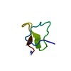
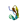
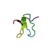
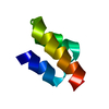
| Title | Crystal structure of the chitin-binding module (CBM18) of a chitinase-like protein from Hevea brasiliensis | ||||||
 Components Components | Class I chitinase | ||||||
 Keywords Keywords | SUGAR BINDING PROTEIN / hevein-like domain / chitin oligomers | ||||||
| Function / homology |  Function and homology information Function and homology informationchitinase activity / chitin catabolic process / chitin binding / defense response to fungus / cell wall macromolecule catabolic process / carbohydrate metabolic process Similarity search - Function | ||||||
| Biological species |  Hevea brasiliensis subsp. brasiliensis (plant) Hevea brasiliensis subsp. brasiliensis (plant) | ||||||
| Method |  X-RAY DIFFRACTION / SYNCHROTRON / MOLECULAR REPLACEMENT / Resolution: 1.602 Å X-RAY DIFFRACTION / SYNCHROTRON / MOLECULAR REPLACEMENT / Resolution: 1.602 Å | ||||||
 Authors Authors | Martinez-Caballero, C.S. / Hermoso, J.A. / Rodriguez-Romero, A. | ||||||
 Citation Citation | Journal: Febs J. / Year: 2014 Title: Comparative study of two GH19 chitinase-like proteins from Hevea brasiliensis, one exhibiting a novel carbohydrate-binding domain. Authors: Martinez-Caballero, S. / Cano-Sanchez, P. / Mares-Mejia, I. / Diaz-Sanchez, A.G. / Macias-Rubalcava, M.L. / Hermoso, J.A. / Rodriguez-Romero, A. | ||||||
| History |
|
- Structure visualization
| Structure viewer | Molecule: MolmilJmol/JSmol |
|---|
- Downloads & links
-Download
| PDBx/mmCIF format | 4mpi.cif.gz | 30.5 KB | Display | PDBx/mmCIF format |
|---|---|---|---|---|
| PDB format | pdb4mpi.ent.gz | 19.9 KB | Display | PDB format |
| PDBx/mmJSON format | 4mpi.json.gz | Tree view | PDBx/mmJSON format | |
| Others |  Other downloads Other downloads |
-Validation report
| Arichive directory | https://data.pdbj.org/pub/pdb/validation_reports/mp/4mpiftp://data.pdbj.org/pub/pdb/validation_reports/mp/4mpi | HTTPS FTP |
|---|
-Related structure data
| Related structure data |  4mstC  1q9bS C: citing same article ( S: Starting model for refinement |
|---|---|
| Similar structure data |


-Links
 PDBj
PDBj- Assembly
| Deposited unit | 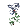
| ||||||||
|---|---|---|---|---|---|---|---|---|---|
| 1 | 
| ||||||||
| 2 | 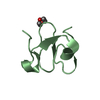
| ||||||||
| Unit cell |
|
-Components
| #1: Protein/peptide | Mass: 4509.008 Da / Num. of mol.: 2 / Fragment: Chitin-binding domain (CBD18, UNP residues 1-43) Source method: isolated from a genetically manipulated source Source: (gene. exp.) Hevea brasiliensis subsp. brasiliensis (plant)Strain: RIMM600 / Tissue: latex and leaves / Gene: Hbchi-L1, laCIC / Plasmid: pET32a / Production host:  #2: Chemical | ChemComp-MES / |   Mass: 195.237 Da / Num. of mol.: 1 / Source method: obtained synthetically / Formula: C6H13NO4S / Comment: pH buffer*YM Mass: 195.237 Da / Num. of mol.: 1 / Source method: obtained synthetically / Formula: C6H13NO4S / Comment: pH buffer*YM#3: Chemical | ChemComp-DIO / |   Mass: 88.105 Da / Num. of mol.: 1 / Source method: obtained synthetically / Formula: C4H8O2 Mass: 88.105 Da / Num. of mol.: 1 / Source method: obtained synthetically / Formula: C4H8O2#4: Water | ChemComp-HOH / |  Mass: 18.015 Da / Num. of mol.: 85 / Source method: isolated from a natural source / Formula: H2O Mass: 18.015 Da / Num. of mol.: 85 / Source method: isolated from a natural source / Formula: H2OHas protein modification | Y | |
|---|
-Experimental details
-Experiment
| Experiment | Method: X-RAY DIFFRACTION / Number of used crystals: 1 |
|---|
- Sample preparation
| Crystal | Density Matthews: 2.4 Å3/Da / Density % sol: 48.67 % |
|---|---|
| Crystal grow | Temperature: 291 K / Method: vapor diffusion, sitting drop / pH: 7 Details: 11 mg/mL protein, 0.1 M MES, pH 7.0, 1.6 M ammonium sulfate, 4% 1,4-dioxane, VAPOR DIFFUSION, SITTING DROP, temperature 291K |
-Data collection
| Diffraction | Mean temperature: 100 K | |||||||||||||||||||||||||||||||||||||||||||||||||||||||||||||||||||||||||||||
|---|---|---|---|---|---|---|---|---|---|---|---|---|---|---|---|---|---|---|---|---|---|---|---|---|---|---|---|---|---|---|---|---|---|---|---|---|---|---|---|---|---|---|---|---|---|---|---|---|---|---|---|---|---|---|---|---|---|---|---|---|---|---|---|---|---|---|---|---|---|---|---|---|---|---|---|---|---|---|
| Diffraction source | Source: SYNCHROTRON / Site: ESRF  / Beamline: ID29 / Wavelength: 0.9791 Å / Beamline: ID29 / Wavelength: 0.9791 Å | |||||||||||||||||||||||||||||||||||||||||||||||||||||||||||||||||||||||||||||
| Detector | Type: DECTRIS PILATUS 6M / Detector: PIXEL / Date: Jun 7, 2012 | |||||||||||||||||||||||||||||||||||||||||||||||||||||||||||||||||||||||||||||
| Radiation | Monochromator: liquid nitrogen cooled channel-cut Si(111) / Protocol: SINGLE WAVELENGTH / Monochromatic (M) / Laue (L): M / Scattering type: x-ray | |||||||||||||||||||||||||||||||||||||||||||||||||||||||||||||||||||||||||||||
| Radiation wavelength | Wavelength: 0.9791 Å / Relative weight: 1 | |||||||||||||||||||||||||||||||||||||||||||||||||||||||||||||||||||||||||||||
| Reflection | Resolution: 1.602→33.355 Å / Num. all: 11176 / Num. obs: 11117 / % possible obs: 99.47 % / Observed criterion σ(F): 1 / Observed criterion σ(I): 1 / Redundancy: 3.5 % / Biso Wilson estimate: 12.65 Å2 / Rmerge(I) obs: 0.037 / Rsym value: 0.037 / Net I/σ(I): 21.13 | |||||||||||||||||||||||||||||||||||||||||||||||||||||||||||||||||||||||||||||
| Reflection shell | Diffraction-ID: 1
|
- Processing
| Software |
| |||||||||||||||||||||||||||||||||||
|---|---|---|---|---|---|---|---|---|---|---|---|---|---|---|---|---|---|---|---|---|---|---|---|---|---|---|---|---|---|---|---|---|---|---|---|---|
| Refinement | Method to determine structure: MOLECULAR REPLACEMENT Starting model: PDB ENTRY 1Q9B Resolution: 1.602→33.355 Å / SU ML: 0.13 / Isotropic thermal model: ISOTROPIC / Cross valid method: THROUGHOUT / σ(F): 1.36 / Phase error: 24.27 / Stereochemistry target values: ML
| |||||||||||||||||||||||||||||||||||
| Solvent computation | Shrinkage radii: 0.9 Å / VDW probe radii: 1.11 Å / Solvent model: FLAT BULK SOLVENT MODEL | |||||||||||||||||||||||||||||||||||
| Refinement step | Cycle: LAST / Resolution: 1.602→33.355 Å
| |||||||||||||||||||||||||||||||||||
| Refine LS restraints |
| |||||||||||||||||||||||||||||||||||
| LS refinement shell |
|
