 Movie
Movie Controller

[English] 日本語
 Yorodumi
- PDB-1q9b: CRYSTAL STRUCTURE ANALYSIS OF Hev b 6.02 (HEVEIN) AT 1.5 ANGSTROM... -
+ Open data

- Basic information
| Entry | Database: PDB / ID: 1q9b | ||||||
|---|---|---|---|---|---|---|---|
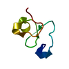
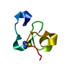
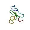
| Title | CRYSTAL STRUCTURE ANALYSIS OF Hev b 6.02 (HEVEIN) AT 1.5 ANGSTROMS RESOLUTION | ||||||
 Components Components | Hevein | ||||||
 Keywords Keywords | ALLERGEN / LECTIN / AGGLUTININ-TOXIN MOTIF | ||||||
| Function / homology |  Function and homology information Function and homology informationchitin binding / RNA nuclease activity / defense response to fungus / defense response to bacterium Similarity search - Function | ||||||
| Biological species |   Hevea brasiliensis (rubber tree) Hevea brasiliensis (rubber tree) | ||||||
| Method |  X-RAY DIFFRACTION / SYNCHROTRON / MOLECULAR REPLACEMENT / Resolution: 1.5 Å X-RAY DIFFRACTION / SYNCHROTRON / MOLECULAR REPLACEMENT / Resolution: 1.5 Å | ||||||
 Authors Authors | Rodriguez-Romero, A. / Hernandez-Santoyo, A. | ||||||
 Citation Citation | Journal: Biochem.Biophys.Res.Commun. / Year: 2004 Title: Insights into a conformational epitope of Hev b 6.02 (hevein). Authors: Reyes-Lopez, C.A. / Hernandez-Santoyo, A. / Pedraza-Escalona, M. / Mendoza, G. / Hernandez-Arana, A. / Rodriguez-Romero, A. #1: Journal: FEBS Lett. / Year: 1991Title: Crystal Structure of Hevein at 2.8 A Resolution Authors: Rodriguez-Romero, A. / Ravichandran, K.G. / Soriano-Garcia, M. | ||||||
| History |
|
- Structure visualization
| Structure viewer | Molecule: MolmilJmol/JSmol |
|---|
- Downloads & links
-Download
| PDBx/mmCIF format | 1q9b.cif.gz | 32.7 KB | Display | PDBx/mmCIF format |
|---|---|---|---|---|
| PDB format | pdb1q9b.ent.gz | 21.8 KB | Display | PDB format |
| PDBx/mmJSON format | 1q9b.json.gz | Tree view | PDBx/mmJSON format | |
| Others |  Other downloads Other downloads |
-Validation report
| Arichive directory | https://data.pdbj.org/pub/pdb/validation_reports/q9/1q9bftp://data.pdbj.org/pub/pdb/validation_reports/q9/1q9b | HTTPS FTP |
|---|
-Related structure data
| Related structure data |  1hevS S: Starting model for refinement |
|---|---|
| Similar structure data |


-Links
 PDBj
PDBj- Assembly
| Deposited unit | 
| ||||||||||
|---|---|---|---|---|---|---|---|---|---|---|---|
| 1 |
| ||||||||||
| Unit cell |
|
-Components
| #1: Protein/peptide | Mass: 4731.166 Da / Num. of mol.: 1 / Source method: isolated from a natural source / Source: (natural) Hevea brasiliensis (rubber tree) / Tissue: LATEX / References: UniProt: P02877 | ||||
|---|---|---|---|---|---|
| #2: Chemical |   Mass: 118.174 Da / Num. of mol.: 2 / Source method: obtained synthetically / Formula: C6H14O2 / Comment: precipitant*YM Mass: 118.174 Da / Num. of mol.: 2 / Source method: obtained synthetically / Formula: C6H14O2 / Comment: precipitant*YM#3: Water | ChemComp-HOH / |  Mass: 18.015 Da / Num. of mol.: 39 / Source method: isolated from a natural source / Formula: H2O Mass: 18.015 Da / Num. of mol.: 39 / Source method: isolated from a natural source / Formula: H2OHas protein modification | Y | |
-Experimental details
-Experiment
| Experiment | Method: X-RAY DIFFRACTION / Number of used crystals: 1 |
|---|
- Sample preparation
| Crystal | Density Matthews: 1.91 Å3/Da / Density % sol: 35.5 % | ||||||||||||||||||
|---|---|---|---|---|---|---|---|---|---|---|---|---|---|---|---|---|---|---|---|
| Crystal grow | Temperature: 291 K / Method: vapor diffusion, sitting drop / pH: 7.1 Details: MPD, Tris-HCl, pH 7.10, VAPOR DIFFUSION, SITTING DROP, temperature 291K | ||||||||||||||||||
| Crystal grow | *PLUS Temperature: 18 ℃ / pH: 7.1 / Method: vapor diffusion, sitting drop | ||||||||||||||||||
| Components of the solutions | *PLUS
|
-Data collection
| Diffraction | Mean temperature: 277 K |
|---|---|
| Diffraction source | Source: SYNCHROTRON / Site: SSRL  / Beamline: BL7-1 / Wavelength: 1.08 / Wavelength: 1.08 Å / Beamline: BL7-1 / Wavelength: 1.08 / Wavelength: 1.08 Å |
| Detector | Type: MARRESEARCH / Detector: IMAGE PLATE / Date: Jan 15, 1999 |
| Radiation | Monochromator: Si / Protocol: SINGLE WAVELENGTH / Monochromatic (M) / Laue (L): M / Scattering type: x-ray |
| Radiation wavelength | Wavelength: 1.08 Å / Relative weight: 1 |
| Reflection | Resolution: 1.5→15 Å / Num. all: 7678 / Num. obs: 7332 / % possible obs: 95.5 % / Redundancy: 4.8 % / Biso Wilson estimate: 13.9 Å2 / Rsym value: 0.05 / Net I/σ(I): 6.7 |
| Reflection shell | Resolution: 1.5→1.53 Å / Redundancy: 4.6 % / Mean I/σ(I) obs: 12.3 / Num. unique all: 635 / Rsym value: 0.046 / % possible all: 97.2 |
| Reflection | *PLUS Highest resolution: 1.5 Å / Lowest resolution: 15 Å / Num. obs: 7113 / Redundancy: 4.7 % / Num. measured all: 57822 / Rmerge(I) obs: 0.05 |
- Processing
| Software |
| |||||||||||||||||||||||||||||||||
|---|---|---|---|---|---|---|---|---|---|---|---|---|---|---|---|---|---|---|---|---|---|---|---|---|---|---|---|---|---|---|---|---|---|---|
| Refinement | Method to determine structure: MOLECULAR REPLACEMENT Starting model: PDB ENTRY 1HEV Resolution: 1.5→9.5 Å / Num. parameters: 3772 / Num. restraintsaints: 4884 / Cross valid method: FREE R / σ(F): 0 / Stereochemistry target values: ENGH AND HUBER Details: THE ALTERNATE CONFORMATIONS EVENTUALLY REDUCED FREE R FROM 21.19-19.20%. INTRODUCTION OF ANISOTROPIC REFINEMENT REDUCED FREE R FROM 19.20- 16.01%. DISCRETELY DISORDERED HETEROGENS: 2, MPD ...Details: THE ALTERNATE CONFORMATIONS EVENTUALLY REDUCED FREE R FROM 21.19-19.20%. INTRODUCTION OF ANISOTROPIC REFINEMENT REDUCED FREE R FROM 19.20- 16.01%. DISCRETELY DISORDERED HETEROGENS: 2, MPD 101 AND MPD 102; DISCRETELY DISORDERED RESIDUES: 6, GLU 1, LEU 11, PRO 13, SER 26, GLU 29, PRO 33. ANISOTROPIC SCALING APPLIED BY THE METHOD OF PARKIN, MOEZZI & HOPE, J.APPL.CRYST.28(1995)53-56
| |||||||||||||||||||||||||||||||||
| Solvent computation | Solvent model: MOEWS & KRETSINGER, J.MOL.BIOL.91(1973)201-228 | |||||||||||||||||||||||||||||||||
| Refine analyze | Num. disordered residues: 8 / Occupancy sum hydrogen: 268 / Occupancy sum non hydrogen: 379 | |||||||||||||||||||||||||||||||||
| Refinement step | Cycle: LAST / Resolution: 1.5→9.5 Å
| |||||||||||||||||||||||||||||||||
| Refine LS restraints |
| |||||||||||||||||||||||||||||||||
| LS refinement shell | Resolution: 1.5→1.53 Å / Num. reflection Rfree: 63 / Num. reflection obs: 635 | |||||||||||||||||||||||||||||||||
| Software | *PLUS Name: SHELXL / Version: 97 / Classification: refinement | |||||||||||||||||||||||||||||||||
| Refinement | *PLUS % reflection Rfree: 10 % / Rfactor Rwork: 0.128 | |||||||||||||||||||||||||||||||||
| Solvent computation | *PLUS | |||||||||||||||||||||||||||||||||
| Displacement parameters | *PLUS | |||||||||||||||||||||||||||||||||
| Refine LS restraints | *PLUS
|
