| Entry | Database: PDB / ID: 4mmr
|
|---|
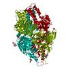
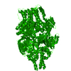
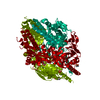
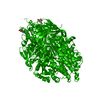
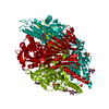
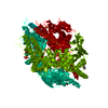
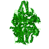
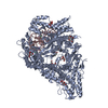
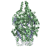
| Title | Crystal Structure of Prefusion-stabilized RSV F Variant Cav1 at pH 9.5 |
|---|
 Components Components | - Fusion glycoprotein F1 fused with Fibritin trimerization domain
- Fusion glycoprotein F2
|
|---|
 Keywords Keywords | VIRAL PROTEIN / fusion / membrane |
|---|
| Function / homology |  Function and homology information Function and homology information
symbiont-mediated induction of syncytium formation / Translation of respiratory syncytial virus mRNAs / RSV-host interactions / Assembly and release of respiratory syncytial virus (RSV) virions / Maturation of hRSV A proteins / Respiratory syncytial virus (RSV) attachment and entry / host cell Golgi membrane / virion component / entry receptor-mediated virion attachment to host cell / fusion of virus membrane with host plasma membrane ...symbiont-mediated induction of syncytium formation / Translation of respiratory syncytial virus mRNAs / RSV-host interactions / Assembly and release of respiratory syncytial virus (RSV) virions / Maturation of hRSV A proteins / Respiratory syncytial virus (RSV) attachment and entry / host cell Golgi membrane / virion component / entry receptor-mediated virion attachment to host cell / fusion of virus membrane with host plasma membrane / viral envelope / symbiont entry into host cell / host cell plasma membrane / virion membrane / identical protein binding / plasma membraneSimilarity search - Function |
|---|
| Biological species |  Human respiratory syncytial virus A2 Human respiratory syncytial virus A2
Enterobacteria phage T4 (virus) |
|---|
| Method |  X-RAY DIFFRACTION / SYNCHROTRON / MOLECULAR REPLACEMENT / Resolution: 3.1 Å X-RAY DIFFRACTION / SYNCHROTRON / MOLECULAR REPLACEMENT / Resolution: 3.1 Å |
|---|
 Authors Authors | Stewart-Jones, G.B.E. / McLellan, J.S. / Joyce, M.G. / Sastry, M. / Yang, Y. / Graham, B.S. / Kwong, P.D. |
|---|
 Citation Citation | Journal: Science / Year: 2013
Title: Structure-based design of a fusion glycoprotein vaccine for respiratory syncytial virus.
Authors: McLellan, J.S. / Chen, M. / Joyce, M.G. / Sastry, M. / Stewart-Jones, G.B. / Yang, Y. / Zhang, B. / Chen, L. / Srivatsan, S. / Zheng, A. / Zhou, T. / Graepel, K.W. / Kumar, A. / Moin, S. / ...Authors: McLellan, J.S. / Chen, M. / Joyce, M.G. / Sastry, M. / Stewart-Jones, G.B. / Yang, Y. / Zhang, B. / Chen, L. / Srivatsan, S. / Zheng, A. / Zhou, T. / Graepel, K.W. / Kumar, A. / Moin, S. / Boyington, J.C. / Chuang, G.Y. / Soto, C. / Baxa, U. / Bakker, A.Q. / Spits, H. / Beaumont, T. / Zheng, Z. / Xia, N. / Ko, S.Y. / Todd, J.P. / Rao, S. / Graham, B.S. / Kwong, P.D. |
|---|
| History | | Deposition | Sep 9, 2013 | Deposition site: RCSB / Processing site: RCSB |
|---|
| Revision 1.0 | Nov 20, 2013 | Provider: repository / Type: Initial release |
|---|
| Revision 1.1 | Jul 26, 2017 | Group: Data collection / Refinement description / Source and taxonomy
Category: diffrn_detector / entity_src_gen / software / Item: _diffrn_detector.detector |
|---|
| Revision 1.2 | Sep 20, 2017 | Group: Database references / Category: struct_ref_seq_dif / Item: _struct_ref_seq_dif.details |
|---|
| Revision 1.3 | Jun 2, 2021 | Group: Database references / Source and taxonomy / Category: entity_src_gen / struct_ref_seq_dif
Item: _entity_src_gen.host_org_common_name / _entity_src_gen.pdbx_host_org_cell_line ..._entity_src_gen.host_org_common_name / _entity_src_gen.pdbx_host_org_cell_line / _entity_src_gen.pdbx_host_org_strain / _struct_ref_seq_dif.details |
|---|
| Revision 1.4 | Nov 20, 2024 | Group: Data collection / Database references / Structure summary
Category: chem_comp_atom / chem_comp_bond ...chem_comp_atom / chem_comp_bond / database_2 / pdbx_entry_details / pdbx_modification_feature
Item: _database_2.pdbx_DOI / _database_2.pdbx_database_accession |
|---|
|
|---|
 Movie
Movie Controller

 Yorodumi
 Open data
 Basic information
 Structure visualization
 Downloads & links
 Other downloads


 PDBj
PDBj


 Assembly
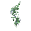
 Homo sapiens (human) / References: UniProt: P03420
 Sample preparation
 / Beamline: 22-ID / Wavelength: 1 Å
 Processing