 Movie
Movie Controller


+ Open data

- Basic information
| Entry | Database: PDB / ID: 4mls | ||||||
|---|---|---|---|---|---|---|---|
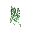
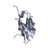
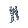
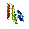
| Title | Crystal structure of the SpyTag and SpyCatcher-deltaN1 complex | ||||||
 Components Components |
| ||||||
 Keywords Keywords | PEPTIDE BINDING PROTEIN / Isopeptide bond / SpyCatcher / Protein engineering | ||||||
| Function / homology |  Function and homology information Function and homology informationFibronectin binding repeat / Fibronectin binding repeat / Uncharacterised domain CHP03934, TQXA / Thioester domain / Thioester domain / Prealbumin-like fold domain / Prealbumin-like fold domain / Immunoglobulin-like fold / Immunoglobulins / Immunoglobulin-like ...Fibronectin binding repeat / Fibronectin binding repeat / Uncharacterised domain CHP03934, TQXA / Thioester domain / Thioester domain / Prealbumin-like fold domain / Prealbumin-like fold domain / Immunoglobulin-like fold / Immunoglobulins / Immunoglobulin-like / Sandwich / Mainly Beta Similarity search - Domain/homology | ||||||
| Biological species |  Streptococcus pyogenes (bacteria) Streptococcus pyogenes (bacteria) | ||||||
| Method |  X-RAY DIFFRACTION / SYNCHROTRON / MOLECULAR REPLACEMENT / Resolution: 1.984 Å X-RAY DIFFRACTION / SYNCHROTRON / MOLECULAR REPLACEMENT / Resolution: 1.984 Å | ||||||
 Authors Authors | Li, L. / Fierer, J.O. / Rapoport, T.A. / Howarth, M. | ||||||
 Citation Citation | Journal: J.Mol.Biol. / Year: 2014 Title: Structural Analysis and Optimization of the Covalent Association between SpyCatcher and a Peptide Tag. Authors: Li, L. / Fierer, J.O. / Rapoport, T.A. / Howarth, M. | ||||||
| History |
|
- Structure visualization
| Structure viewer | Molecule: MolmilJmol/JSmol |
|---|
- Downloads & links
-Download
| PDBx/mmCIF format | 4mls.cif.gz | 51.1 KB | Display | PDBx/mmCIF format |
|---|---|---|---|---|
| PDB format | pdb4mls.ent.gz | 36.6 KB | Display | PDB format |
| PDBx/mmJSON format | 4mls.json.gz | Tree view | PDBx/mmJSON format | |
| Others |  Other downloads Other downloads |
-Validation report
| Arichive directory | https://data.pdbj.org/pub/pdb/validation_reports/ml/4mlsftp://data.pdbj.org/pub/pdb/validation_reports/ml/4mls | HTTPS FTP |
|---|
-Related structure data
| Related structure data |  4mliC 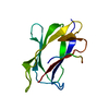 2x5pS C: citing same article ( S: Starting model for refinement |
|---|---|
| Similar structure data |


-Links
 PDBj
PDBj- Assembly
| Deposited unit | 
| ||||||||
|---|---|---|---|---|---|---|---|---|---|
| 1 |
| ||||||||
| Unit cell |
| ||||||||
| Components on special symmetry positions |
|
-Components
| #1: Protein | Mass: 10192.143 Da / Num. of mol.: 1 / Fragment: UNP residues 461-552 / Mutation: E473I, Y508M Source method: isolated from a genetically manipulated source Source: (gene. exp.) Streptococcus pyogenes (bacteria) / Gene: CnaB2, fba2 / Plasmid: pDEST14 / Production host: |
|---|---|
| #2: Protein/peptide | Mass: 1475.773 Da / Num. of mol.: 1 / Source method: obtained synthetically / Details: Chemically synthesized |
| #3: Water | ChemComp-HOH /  Mass: 18.015 Da / Num. of mol.: 61 / Source method: isolated from a natural source / Formula: H2O Mass: 18.015 Da / Num. of mol.: 61 / Source method: isolated from a natural source / Formula: H2O |
| Has protein modification | Y |
-Experimental details
-Experiment
| Experiment | Method: X-RAY DIFFRACTION / Number of used crystals: 1 |
|---|
- Sample preparation
| Crystal | Density Matthews: 1.83 Å3/Da / Density % sol: 32.77 % |
|---|---|
| Crystal grow | Temperature: 295 K / Method: vapor diffusion, hanging drop / pH: 4.5 Details: Sodium Acetate 0.1M pH4.5, NaCl 200mM, PEG3350 30%, Glycerol 5%, VAPOR DIFFUSION, HANGING DROP, temperature 295KK |
-Data collection
| Diffraction | Mean temperature: 100 K |
|---|---|
| Diffraction source | Source: SYNCHROTRON / Site: APS  / Beamline: 24-ID-C / Wavelength: 0.9792 Å / Beamline: 24-ID-C / Wavelength: 0.9792 Å |
| Detector | Type: PSI PILATUS 6M / Detector: PIXEL / Date: Oct 11, 2012 |
| Radiation | Monochromator: Si(111) / Protocol: SINGLE WAVELENGTH / Monochromatic (M) / Laue (L): M / Scattering type: x-ray |
| Radiation wavelength | Wavelength: 0.9792 Å / Relative weight: 1 |
| Reflection | Resolution: 1.984→50 Å / Num. all: 5858 / Num. obs: 5530 / % possible obs: 94.4 % / Observed criterion σ(F): 0 / Observed criterion σ(I): -3 / Redundancy: 3 % / Biso Wilson estimate: 24.7 Å2 / Rmerge(I) obs: 0.117 / Rsym value: 0.117 / Net I/σ(I): 12.6 |
| Reflection shell | Resolution: 2→2.03 Å / Redundancy: 2.9 % / Rmerge(I) obs: 0.362 / Mean I/σ(I) obs: 5.2 / Num. unique all: 281 / Rsym value: 0.362 / % possible all: 94.6 |
- Processing
| Software |
| |||||||||||||||||||||||||||||||||||||||||||||||||||||||||||||||||||||||||||
|---|---|---|---|---|---|---|---|---|---|---|---|---|---|---|---|---|---|---|---|---|---|---|---|---|---|---|---|---|---|---|---|---|---|---|---|---|---|---|---|---|---|---|---|---|---|---|---|---|---|---|---|---|---|---|---|---|---|---|---|---|---|---|---|---|---|---|---|---|---|---|---|---|---|---|---|---|
| Refinement | Method to determine structure: MOLECULAR REPLACEMENT Starting model: PDB ENTRY 2X5P Resolution: 1.984→39.394 Å / SU ML: 0.3 / σ(F): 1.41 / Phase error: 31.25 / Stereochemistry target values: ML
| |||||||||||||||||||||||||||||||||||||||||||||||||||||||||||||||||||||||||||
| Solvent computation | Shrinkage radii: 0.9 Å / VDW probe radii: 1.11 Å / Solvent model: FLAT BULK SOLVENT MODEL | |||||||||||||||||||||||||||||||||||||||||||||||||||||||||||||||||||||||||||
| Refinement step | Cycle: LAST / Resolution: 1.984→39.394 Å
| |||||||||||||||||||||||||||||||||||||||||||||||||||||||||||||||||||||||||||
| Refine LS restraints |
| |||||||||||||||||||||||||||||||||||||||||||||||||||||||||||||||||||||||||||
| LS refinement shell |
| |||||||||||||||||||||||||||||||||||||||||||||||||||||||||||||||||||||||||||
| Refinement TLS params. | Method: refined / Refine-ID: X-RAY DIFFRACTION
| |||||||||||||||||||||||||||||||||||||||||||||||||||||||||||||||||||||||||||
| Refinement TLS group |
|
