 Movie
Movie Controller

[English] 日本語
 Yorodumi
- PDB-4mln: Crystal of PhnZ bound to (R)-2-amino-1-hydroxyethylphosphonic acid -
+ Open data

- Basic information
| Entry | Database: PDB / ID: 4mln | ||||||
|---|---|---|---|---|---|---|---|
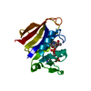
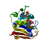
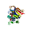
| Title | Crystal of PhnZ bound to (R)-2-amino-1-hydroxyethylphosphonic acid | ||||||
 Components Components | Predicted HD phosphohydrolase PhnZ | ||||||
 Keywords Keywords | HYDROLASE / Structural Genomics / Montreal-Kingston Bacterial Structural Genomics Initiative / BSGI / all alpha / Carbon-phosphorus bond cleavage | ||||||
| Function / homology |  Function and homology information Function and homology information2-amino-1-hydroxyethylphosphonate dioxygenase (glycine-forming) / oxidoreductase activity / hydrolase activity / metal ion binding Similarity search - Function | ||||||
| Biological species |  uncultured bacterium HF130_AEPn_1 (environmental samples) uncultured bacterium HF130_AEPn_1 (environmental samples) | ||||||
| Method |  X-RAY DIFFRACTION / SYNCHROTRON / MOLECULAR REPLACEMENT / Resolution: 2.1 Å X-RAY DIFFRACTION / SYNCHROTRON / MOLECULAR REPLACEMENT / Resolution: 2.1 Å | ||||||
 Authors Authors | van Staalduinen, L.M. / McSorley, F.R. / Zechel, D.L. / Jia, Z. / Montreal-Kingston Bacterial Structural Genomics Initiative (BSGI) | ||||||
 Citation Citation | Journal: Proc.Natl.Acad.Sci.USA / Year: 2014 Title: Crystal structure of PhnZ in complex with substrate reveals a di-iron oxygenase mechanism for catabolism of organophosphonates. Authors: van Staalduinen, L.M. / McSorley, F.R. / Schiessl, K. / Seguin, J. / Wyatt, P.B. / Hammerschmidt, F. / Zechel, D.L. / Jia, Z. | ||||||
| History |
|
- Structure visualization
| Structure viewer | Molecule: MolmilJmol/JSmol |
|---|
- Downloads & links
-Download
| PDBx/mmCIF format | 4mln.cif.gz | 87.8 KB | Display | PDBx/mmCIF format |
|---|---|---|---|---|
| PDB format | pdb4mln.ent.gz | 66.1 KB | Display | PDB format |
| PDBx/mmJSON format | 4mln.json.gz | Tree view | PDBx/mmJSON format | |
| Others |  Other downloads Other downloads |
-Validation report
| Arichive directory | https://data.pdbj.org/pub/pdb/validation_reports/ml/4mlnftp://data.pdbj.org/pub/pdb/validation_reports/ml/4mln | HTTPS FTP |
|---|
-Related structure data
| Related structure data |  4mlmSC S: Starting model for refinement C: citing same article ( |
|---|---|
| Similar structure data |


-Links
 PDBj
PDBj
- Assembly
| Deposited unit | 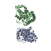
| ||||||||
|---|---|---|---|---|---|---|---|---|---|
| 1 | 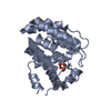
| ||||||||
| 2 | 
| ||||||||
| Unit cell |
|
-Components
| #1: Protein | Mass: 22446.133 Da / Num. of mol.: 2 Source method: isolated from a genetically manipulated source Source: (gene. exp.) uncultured bacterium HF130_AEPn_1 (environmental samples)Gene: ALOHA_HF130_AEPn_1_06c / Plasmid: pJExpress / Production host: #2: Chemical | ChemComp-FE /   Mass: 55.845 Da / Num. of mol.: 4 / Source method: obtained synthetically / Formula: Fe Mass: 55.845 Da / Num. of mol.: 4 / Source method: obtained synthetically / Formula: Fe#3: Chemical |   Mass: 141.063 Da / Num. of mol.: 2 / Source method: obtained synthetically / Formula: C2H8NO4P Mass: 141.063 Da / Num. of mol.: 2 / Source method: obtained synthetically / Formula: C2H8NO4P#4: Water | ChemComp-HOH / |  Mass: 18.015 Da / Num. of mol.: 138 / Source method: isolated from a natural source / Formula: H2O Mass: 18.015 Da / Num. of mol.: 138 / Source method: isolated from a natural source / Formula: H2O |
|---|
-Experimental details
-Experiment
| Experiment | Method: X-RAY DIFFRACTION |
|---|
- Sample preparation
| Crystal | Density Matthews: 2.11 Å3/Da / Density % sol: 41.63 % |
|---|---|
| Crystal grow | Temperature: 294 K / Method: vapor diffusion, hanging drop / pH: 6.5 Details: 20 % PEG 5000 MME, 0.1 M Bis-Tris pH 6.5, VAPOR DIFFUSION, HANGING DROP, temperature 294K |
-Data collection
| Diffraction | Mean temperature: 100 K |
|---|---|
| Diffraction source | Source: SYNCHROTRON / Site: APS  / Beamline: 23-ID-B / Wavelength: 1.0332 Å / Beamline: 23-ID-B / Wavelength: 1.0332 Å |
| Detector | Type: MARMOSAIC 300 mm CCD / Detector: CCD / Date: Aug 9, 2012 |
| Radiation | Monochromator: double crystal / Protocol: SINGLE WAVELENGTH / Monochromatic (M) / Laue (L): M / Scattering type: x-ray |
| Radiation wavelength | Wavelength: 1.0332 Å / Relative weight: 1 |
| Reflection | Resolution: 2.1→20 Å / Num. all: 22754 / Num. obs: 22671 / % possible obs: 99.6 % / Observed criterion σ(F): 1 / Observed criterion σ(I): 1 / Redundancy: 7.3 % / Rsym value: 0.111 / Net I/σ(I): 12 |
| Reflection shell | Resolution: 2.1→2.2 Å / % possible all: 99.7 |
- Processing
| Software |
| |||||||||||||||||||||||||||||||||||||||||||||||||||||||||||||||
|---|---|---|---|---|---|---|---|---|---|---|---|---|---|---|---|---|---|---|---|---|---|---|---|---|---|---|---|---|---|---|---|---|---|---|---|---|---|---|---|---|---|---|---|---|---|---|---|---|---|---|---|---|---|---|---|---|---|---|---|---|---|---|---|---|
| Refinement | Method to determine structure: MOLECULAR REPLACEMENT Starting model: PDB Entry 4MLM Resolution: 2.1→19.9 Å / SU ML: 0.26 / σ(F): 2 / Phase error: 24.23 / Stereochemistry target values: ML
| |||||||||||||||||||||||||||||||||||||||||||||||||||||||||||||||
| Solvent computation | Shrinkage radii: 0.9 Å / VDW probe radii: 1.11 Å / Solvent model: FLAT BULK SOLVENT MODEL | |||||||||||||||||||||||||||||||||||||||||||||||||||||||||||||||
| Refinement step | Cycle: LAST / Resolution: 2.1→19.9 Å
| |||||||||||||||||||||||||||||||||||||||||||||||||||||||||||||||
| Refine LS restraints |
| |||||||||||||||||||||||||||||||||||||||||||||||||||||||||||||||
| LS refinement shell |
|
