 Movie
Movie Controller


+ Open data

- Basic information
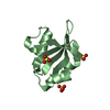
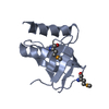
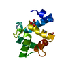
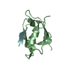
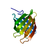
| Entry | Database: PDB / ID: 4ml2 | ||||||
|---|---|---|---|---|---|---|---|
| Title | Crystal structure of wild-type YafQ | ||||||
 Components Components | mRNA interferase YafQ | ||||||
 Keywords Keywords | TOXIN / antitoxin | ||||||
| Function / homology |  Function and homology information Function and homology informationsingle-species biofilm formation / regulation of growth / mRNA catabolic process / translational termination / RNA endonuclease activity / RNA endonuclease activity producing 3'-phosphomonoesters, hydrolytic mechanism / ribosome binding / Hydrolases; Acting on ester bonds / response to antibiotic / negative regulation of DNA-templated transcription ...single-species biofilm formation / regulation of growth / mRNA catabolic process / translational termination / RNA endonuclease activity / RNA endonuclease activity producing 3'-phosphomonoesters, hydrolytic mechanism / ribosome binding / Hydrolases; Acting on ester bonds / response to antibiotic / negative regulation of DNA-templated transcription / DNA-templated transcription / DNA binding / RNA binding Similarity search - Function | ||||||
| Biological species |  | ||||||
| Method |  X-RAY DIFFRACTION / SYNCHROTRON / MOLECULAR REPLACEMENT / Resolution: 1.5 Å X-RAY DIFFRACTION / SYNCHROTRON / MOLECULAR REPLACEMENT / Resolution: 1.5 Å | ||||||
 Authors Authors | Liang, Y.J. / Gao, Z.Q. / Liu, Q.S. / Dong, Y.H. | ||||||
 Citation Citation | Journal: J.Biol.Chem. / Year: 2014 Title: Structural and Functional Characterization of Escherichia coli Toxin-Antitoxin Complex DinJ-YafQ Authors: Liang, Y. / Gao, Z. / Wang, F. / Zhang, Y. / Dong, Y. / Liu, Q. | ||||||
| History |
|
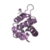
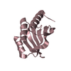
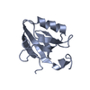
- Structure visualization
| Structure viewer | Molecule: MolmilJmol/JSmol |
|---|
- Downloads & links
-Download
| PDBx/mmCIF format | 4ml2.cif.gz | 38.5 KB | Display | PDBx/mmCIF format |
|---|---|---|---|---|
| PDB format | pdb4ml2.ent.gz | 25.4 KB | Display | PDB format |
| PDBx/mmJSON format | 4ml2.json.gz | Tree view | PDBx/mmJSON format | |
| Others |  Other downloads Other downloads |
-Validation report
| Arichive directory | https://data.pdbj.org/pub/pdb/validation_reports/ml/4ml2ftp://data.pdbj.org/pub/pdb/validation_reports/ml/4ml2 | HTTPS FTP |
|---|
-Related structure data
| Related structure data |  4ml0C  4mmgSC  4mmjC C: citing same article ( S: Starting model for refinement |
|---|---|
| Similar structure data |

-Links
 PDBj
PDBj- Assembly
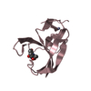
| Deposited unit | 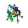
| ||||||||
|---|---|---|---|---|---|---|---|---|---|
| 1 |
| ||||||||
| Unit cell |
|
-Components
| #1: Protein | Mass: 11110.871 Da / Num. of mol.: 1 Source method: isolated from a genetically manipulated source Source: (gene. exp.) References: UniProt: Q47149, Hydrolases; Acting on ester bonds | ||
|---|---|---|---|
| #2: Chemical | ChemComp-SO4 /   Mass: 96.063 Da / Num. of mol.: 5 / Source method: obtained synthetically / Formula: SO4 Mass: 96.063 Da / Num. of mol.: 5 / Source method: obtained synthetically / Formula: SO4#3: Water | ChemComp-HOH / |  Mass: 18.015 Da / Num. of mol.: 217 / Source method: isolated from a natural source / Formula: H2O Mass: 18.015 Da / Num. of mol.: 217 / Source method: isolated from a natural source / Formula: H2O |
-Experimental details
-Experiment
| Experiment | Method: X-RAY DIFFRACTION / Number of used crystals: 1 |
|---|
- Sample preparation
| Crystal | Density Matthews: 3.13 Å3/Da / Density % sol: 60.71 % |
|---|---|
| Crystal grow | Temperature: 293 K / Method: vapor diffusion, hanging drop / pH: 7 Details: 0.2M (NH4)2SO4, 30% PEG8000, pH 7.0, VAPOR DIFFUSION, HANGING DROP, temperature 293K |
-Data collection
| Diffraction | Mean temperature: 100 K |
|---|---|
| Diffraction source | Source: SYNCHROTRON / Site: BSRF  / Beamline: 1W2B / Wavelength: 0.9792 Å / Beamline: 1W2B / Wavelength: 0.9792 Å |
| Detector | Type: MAR CCD 165 mm / Detector: CCD / Date: Jul 19, 2012 |
| Radiation | Monochromator: double crystal monochromator / Protocol: SINGLE WAVELENGTH / Monochromatic (M) / Laue (L): M / Scattering type: x-ray |
| Radiation wavelength | Wavelength: 0.9792 Å / Relative weight: 1 |
| Reflection | Resolution: 1.5→50 Å / Num. all: 22814 / Num. obs: 22754 / % possible obs: 99.7 % / Observed criterion σ(F): 0 / Observed criterion σ(I): 0 / Redundancy: 10 % / Rmerge(I) obs: 0.062 / Net I/σ(I): 72.5 |
| Reflection shell | Resolution: 1.5→1.53 Å / Redundancy: 6.9 % / Rmerge(I) obs: 0.167 / Mean I/σ(I) obs: 16.3 / Num. unique all: 1104 / % possible all: 100 |
- Processing
| Software |
| |||||||||||||||||||||||||||||||||||||||||||||
|---|---|---|---|---|---|---|---|---|---|---|---|---|---|---|---|---|---|---|---|---|---|---|---|---|---|---|---|---|---|---|---|---|---|---|---|---|---|---|---|---|---|---|---|---|---|---|
| Refinement | Method to determine structure: MOLECULAR REPLACEMENT Starting model: 4MMG Resolution: 1.5→28.79 Å / SU ML: 0.17 / σ(F): 1.35 / Phase error: 15.21 / Stereochemistry target values: ML
| |||||||||||||||||||||||||||||||||||||||||||||
| Solvent computation | Shrinkage radii: 0.86 Å / VDW probe radii: 1.1 Å / Solvent model: FLAT BULK SOLVENT MODEL / Bsol: 45.103 Å2 / ksol: 0.4 e/Å3 | |||||||||||||||||||||||||||||||||||||||||||||
| Displacement parameters |
| |||||||||||||||||||||||||||||||||||||||||||||
| Refinement step | Cycle: LAST / Resolution: 1.5→28.79 Å
| |||||||||||||||||||||||||||||||||||||||||||||
| Refine LS restraints |
| |||||||||||||||||||||||||||||||||||||||||||||
| LS refinement shell | Refine-ID: X-RAY DIFFRACTION / Total num. of bins used: 8 / % reflection obs: 100 %
|
