 Movie
Movie Controller

[English] 日本語
 Yorodumi
- PDB-4mdy: Crystal structure of periplasmic solute binding protein from Myco... -
+ Open data

- Basic information
| Entry | Database: PDB / ID: 4mdy | ||||||
|---|---|---|---|---|---|---|---|
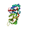
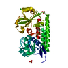
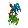
| Title | Crystal structure of periplasmic solute binding protein from Mycobacterium smegmatis str. MC2 155 | ||||||
 Components Components | Periplasmic binding protein | ||||||
 Keywords Keywords | TRANSPORT PROTEIN / MCSG / STRUCTURAL GENOMICS / PSI-BIOLOGY / MIDWEST CENTER FOR STRUCTURAL GENOMICS / Protein Structure Initiative / ABC transporter system / solute binding protein | ||||||
| Function / homology |  Function and homology information Function and homology informationABC transporter periplasmic binding domain / Periplasmic binding protein / Iron siderophore/cobalamin periplasmic-binding domain profile. / Nitrogenase molybdenum iron protein domain / Twin arginine translocation (Tat) signal profile. / Twin-arginine translocation pathway, signal sequence / Rossmann fold / 3-Layer(aba) Sandwich / Alpha Beta Similarity search - Domain/homology | ||||||
| Biological species |  Mycobacterium smegmatis (bacteria) Mycobacterium smegmatis (bacteria) | ||||||
| Method |  X-RAY DIFFRACTION / SYNCHROTRON / SAD / Resolution: 1.78 Å X-RAY DIFFRACTION / SYNCHROTRON / SAD / Resolution: 1.78 Å | ||||||
 Authors Authors | Chang, C. / Wu, R. / Endres, M. / Joachimiak, A. / Midwest Center for Structural Genomics (MCSG) | ||||||
 Citation Citation | Journal: To be Published Title: Crystal structure of periplasmic solute binding protein from Mycobacterium smegmatis str. MC2 155 Authors: Chang, C. / Wu, R. / Endres, M. / Joachimiak, A. | ||||||
| History |
|
- Structure visualization
| Structure viewer | Molecule: MolmilJmol/JSmol |
|---|
- Downloads & links
-Download
| PDBx/mmCIF format | 4mdy.cif.gz | 125.8 KB | Display | PDBx/mmCIF format |
|---|---|---|---|---|
| PDB format | pdb4mdy.ent.gz | 102.2 KB | Display | PDB format |
| PDBx/mmJSON format | 4mdy.json.gz | Tree view | PDBx/mmJSON format | |
| Others |  Other downloads Other downloads |
-Validation report
| Summary document | 4mdy_validation.pdf.gz | 425.9 KB | Display | wwPDB validaton report |
|---|---|---|---|---|
| Full document | 4mdy_full_validation.pdf.gz | 426.1 KB | Display | |
| Data in XML | 4mdy_validation.xml.gz | 13.8 KB | Display | |
| Data in CIF | 4mdy_validation.cif.gz | 19.8 KB | Display | |
| Arichive directory | https://data.pdbj.org/pub/pdb/validation_reports/md/4mdyftp://data.pdbj.org/pub/pdb/validation_reports/md/4mdy | HTTPS FTP |
-Related structure data
| Similar structure data | |
|---|---|
| Other databases |


-Links
 PDBj
PDBj

- Assembly
| Deposited unit | 
| ||||||||
|---|---|---|---|---|---|---|---|---|---|
| 1 |
| ||||||||
| Unit cell |
|
-Components
| #1: Protein | Mass: 31493.086 Da / Num. of mol.: 1 Source method: isolated from a genetically manipulated source Source: (gene. exp.) Mycobacterium smegmatis (bacteria) / Strain: MC2 155 / Gene: MSMEG_0438, MSMEI_0427 / Plasmid: pMCSG68 / Production host: |
|---|---|
| #2: Chemical | ChemComp-PEG /   Mass: 106.120 Da / Num. of mol.: 1 / Source method: obtained synthetically / Formula: C4H10O3 Mass: 106.120 Da / Num. of mol.: 1 / Source method: obtained synthetically / Formula: C4H10O3 |
| #3: Water | ChemComp-HOH /  Mass: 18.015 Da / Num. of mol.: 199 / Source method: isolated from a natural source / Formula: H2O Mass: 18.015 Da / Num. of mol.: 199 / Source method: isolated from a natural source / Formula: H2O |
-Experimental details
-Experiment
| Experiment | Method: X-RAY DIFFRACTION / Number of used crystals: 1 |
|---|
- Sample preparation
| Crystal | Density Matthews: 2.04 Å3/Da / Density % sol: 39.69 % |
|---|---|
| Crystal grow | Temperature: 289 K / Method: vapor diffusion, sitting drop / pH: 4.2 Details: 0.2M Sodium Citrate, 0.1 M Phosphate-citrate, 20 % PEG 8000, pH 4.2, VAPOR DIFFUSION, SITTING DROP, temperature 289K |
-Data collection
| Diffraction | Mean temperature: 100 K |
|---|---|
| Diffraction source | Source: SYNCHROTRON / Site: APS  / Beamline: 19-ID / Wavelength: 0.97899 Å / Beamline: 19-ID / Wavelength: 0.97899 Å |
| Detector | Type: ADSC QUANTUM 315r / Detector: CCD / Date: Dec 16, 2012 |
| Radiation | Monochromator: Si(111) double crystal / Protocol: SINGLE WAVELENGTH / Monochromatic (M) / Laue (L): M / Scattering type: x-ray |
| Radiation wavelength | Wavelength: 0.97899 Å / Relative weight: 1 |
| Reflection | Resolution: 1.78→50 Å / Num. all: 25588 / Num. obs: 24908 / % possible obs: 97.3 % / Observed criterion σ(I): -3 / Redundancy: 6.9 % / Rmerge(I) obs: 0.091 / Net I/σ(I): 23.1 |
| Reflection shell | Resolution: 1.78→1.81 Å / Redundancy: 6.1 % / Rmerge(I) obs: 0.992 / Mean I/σ(I) obs: 1.92 / Num. unique all: 1170 / % possible all: 97.3 |
- Processing
| Software |
| |||||||||||||||||||||||||||||||||||||||||||||||||||||||||||||||||||||||||||
|---|---|---|---|---|---|---|---|---|---|---|---|---|---|---|---|---|---|---|---|---|---|---|---|---|---|---|---|---|---|---|---|---|---|---|---|---|---|---|---|---|---|---|---|---|---|---|---|---|---|---|---|---|---|---|---|---|---|---|---|---|---|---|---|---|---|---|---|---|---|---|---|---|---|---|---|---|
| Refinement | Method to determine structure: SAD / Resolution: 1.78→31.82 Å / Cor.coef. Fo:Fc: 0.971 / Cor.coef. Fo:Fc free: 0.948 / Occupancy max: 1 / Occupancy min: 0.5 / SU B: 5.729 / SU ML: 0.081 / Cross valid method: THROUGHOUT / σ(F): 0 / ESU R Free: 0.128 Stereochemistry target values: MAXIMUM LIKELIHOOD WITH PHASES Details: HYDROGENS HAVE BEEN ADDED IN THE RIDING POSITIONS U VALUES : REFINED INDIVIDUALLY
| |||||||||||||||||||||||||||||||||||||||||||||||||||||||||||||||||||||||||||
| Solvent computation | Ion probe radii: 0.8 Å / Shrinkage radii: 0.8 Å / VDW probe radii: 1.2 Å / Solvent model: MASK | |||||||||||||||||||||||||||||||||||||||||||||||||||||||||||||||||||||||||||
| Displacement parameters | Biso max: 78.07 Å2 / Biso mean: 25.3141 Å2 / Biso min: 11.72 Å2
| |||||||||||||||||||||||||||||||||||||||||||||||||||||||||||||||||||||||||||
| Refinement step | Cycle: LAST / Resolution: 1.78→31.82 Å
| |||||||||||||||||||||||||||||||||||||||||||||||||||||||||||||||||||||||||||
| Refine LS restraints |
| |||||||||||||||||||||||||||||||||||||||||||||||||||||||||||||||||||||||||||
| LS refinement shell | Resolution: 1.779→1.825 Å / Total num. of bins used: 20
|
