 Movie
Movie Controller


+ Open data

- Basic information
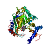
| Entry | Database: PDB / ID: 4mc8 | ||||||
|---|---|---|---|---|---|---|---|
| Title | Hedycaryol synthase in complex with HEPES | ||||||
 Components Components | Putative sesquiterpene cyclase | ||||||
 Keywords Keywords | LYASE / cyclase / terpenoid / Terpene alpha domain class I / helix break / helix dipol | ||||||
| Function / homology |  Function and homology information Function and homology information(2Z,6E)-hedycaryol synthase / terpene synthase activity / terpenoid biosynthetic process / metal ion binding Similarity search - Function | ||||||
| Biological species |  Kitasatospora setae (bacteria) Kitasatospora setae (bacteria) | ||||||
| Method |  X-RAY DIFFRACTION / SYNCHROTRON / MOLECULAR REPLACEMENT / Resolution: 1.9 Å X-RAY DIFFRACTION / SYNCHROTRON / MOLECULAR REPLACEMENT / Resolution: 1.9 Å | ||||||
 Authors Authors | Baer, P. / Rabe, P. / Cirton, C. / Oliveira Mann, C. / Kaufmann, N. / Groll, M. / Dickschat, J. | ||||||
 Citation Citation | Journal: Chembiochem / Year: 2014 Title: Hedycaryol synthase in complex with nerolidol reveals terpene cyclase mechanism. Authors: Baer, P. / Rabe, P. / Citron, C.A. / de Oliveira Mann, C.C. / Kaufmann, N. / Groll, M. / Dickschat, J.S. | ||||||
| History |
|
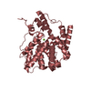
- Structure visualization
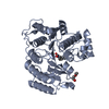
| Structure viewer | Molecule: MolmilJmol/JSmol |
|---|
- Downloads & links
-Download
| PDBx/mmCIF format | 4mc8.cif.gz | 143.8 KB | Display | PDBx/mmCIF format |
|---|---|---|---|---|
| PDB format | pdb4mc8.ent.gz | 112.6 KB | Display | PDB format |
| PDBx/mmJSON format | 4mc8.json.gz | Tree view | PDBx/mmJSON format | |
| Others |  Other downloads Other downloads |
-Validation report
| Arichive directory | https://data.pdbj.org/pub/pdb/validation_reports/mc/4mc8ftp://data.pdbj.org/pub/pdb/validation_reports/mc/4mc8 | HTTPS FTP |
|---|
-Related structure data
| Related structure data |  4mc0C  4mc3SC C: citing same article ( S: Starting model for refinement |
|---|---|
| Similar structure data |


-Links
 PDBj
PDBj
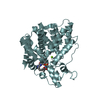
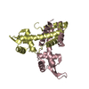
- Assembly
| Deposited unit | 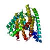
| ||||||||
|---|---|---|---|---|---|---|---|---|---|
| 1 |
| ||||||||
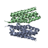
| Unit cell |
|
-Components
| #1: Protein | Mass: 38701.473 Da / Num. of mol.: 1 Source method: isolated from a genetically manipulated source Source: (gene. exp.) Kitasatospora setae (bacteria)Strain: ATCC 33774 / DSM 43861 / JCM 3304 / KCC A-0304 / NBRC 14216 / KM-6054 Gene: KSE_00200t, KSE_76540t / Plasmid: pET28c / Production host: References: UniProt: E4MYY0, Lyases; Carbon-oxygen lyases; Acting on phosphates |
|---|---|
| #2: Chemical | ChemComp-EPE /   Mass: 238.305 Da / Num. of mol.: 1 / Source method: obtained synthetically / Formula: C8H18N2O4S / Comment: pH buffer*YM Mass: 238.305 Da / Num. of mol.: 1 / Source method: obtained synthetically / Formula: C8H18N2O4S / Comment: pH buffer*YM |
| #3: Water | ChemComp-HOH /  Mass: 18.015 Da / Num. of mol.: 208 / Source method: isolated from a natural source / Formula: H2O Mass: 18.015 Da / Num. of mol.: 208 / Source method: isolated from a natural source / Formula: H2O |
-Experimental details
-Experiment
| Experiment | Method: X-RAY DIFFRACTION / Number of used crystals: 1 |
|---|
- Sample preparation
| Crystal | Density Matthews: 3.31 Å3/Da / Density % sol: 62.82 % |
|---|---|
| Crystal grow | Temperature: 293 K / Method: vapor diffusion, hanging drop / pH: 7.5 Details: 0.1 M, HEPES, 20 mM MgCl2, 21% Sodium polyacrylate 5100, pH 7.5, VAPOR DIFFUSION, HANGING DROP, temperature 293K |
-Data collection
| Diffraction | Mean temperature: 100 K |
|---|---|
| Diffraction source | Source: SYNCHROTRON / Site: SLS  / Beamline: X06SA / Wavelength: 1 Å / Beamline: X06SA / Wavelength: 1 Å |
| Detector | Type: PSI PILATUS 6M / Detector: PIXEL / Date: Feb 23, 2013 |
| Radiation | Monochromator: LN2 cooled fixed-exit. Si(111) monochromator / Protocol: SINGLE WAVELENGTH / Monochromatic (M) / Laue (L): M / Scattering type: x-ray |
| Radiation wavelength | Wavelength: 1 Å / Relative weight: 1 |
| Reflection | Resolution: 1.9→38.71 Å / Num. all: 42526 / Num. obs: 42016 / % possible obs: 98.8 % / Observed criterion σ(F): 2 / Observed criterion σ(I): 2 / Redundancy: 6.2 % / Rmerge(I) obs: 0.087 / Net I/σ(I): 10.4 |
| Reflection shell | Resolution: 1.9→2 Å / Rmerge(I) obs: 5.94 / Mean I/σ(I) obs: 2.1 / % possible all: 93 |
- Processing
| Software |
| ||||||||||||||||||||||||||||||||||||||||||||||||||||||||||||
|---|---|---|---|---|---|---|---|---|---|---|---|---|---|---|---|---|---|---|---|---|---|---|---|---|---|---|---|---|---|---|---|---|---|---|---|---|---|---|---|---|---|---|---|---|---|---|---|---|---|---|---|---|---|---|---|---|---|---|---|---|---|
| Refinement | Method to determine structure: MOLECULAR REPLACEMENT Starting model: PDB entry 4MC3 Resolution: 1.9→38.71 Å / Cor.coef. Fo:Fc: 0.966 / Cor.coef. Fo:Fc free: 0.956 / SU B: 8.646 / SU ML: 0.105 / Cross valid method: THROUGHOUT / σ(F): 2 / ESU R: 0.14 / ESU R Free: 0.108 / Stereochemistry target values: MAXIMUM LIKELIHOOD
| ||||||||||||||||||||||||||||||||||||||||||||||||||||||||||||
| Solvent computation | Ion probe radii: 0.8 Å / Shrinkage radii: 0.8 Å / VDW probe radii: 1.2 Å / Solvent model: MASK | ||||||||||||||||||||||||||||||||||||||||||||||||||||||||||||
| Displacement parameters | Biso mean: 65.959 Å2
| ||||||||||||||||||||||||||||||||||||||||||||||||||||||||||||
| Refinement step | Cycle: LAST / Resolution: 1.9→38.71 Å
| ||||||||||||||||||||||||||||||||||||||||||||||||||||||||||||
| Refine LS restraints |
| ||||||||||||||||||||||||||||||||||||||||||||||||||||||||||||
| LS refinement shell | Resolution: 1.9→1.952 Å / Total num. of bins used: 20
| ||||||||||||||||||||||||||||||||||||||||||||||||||||||||||||
| Refinement TLS params. | Method: refined / Origin x: -16.6239 Å / Origin y: 34.4506 Å / Origin z: 7.6602 Å
|
