 Movie
Movie Controller

[English] 日本語
 Yorodumi
- PDB-4m7v: Dihydrofolate reductase from Enterococcus faecalis complexed with... -
+ Open data

- Basic information
| Entry | Database: PDB / ID: 4m7v | ||||||
|---|---|---|---|---|---|---|---|
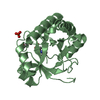
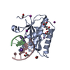
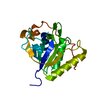
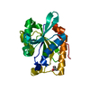
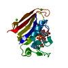
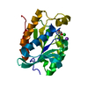
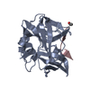
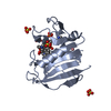
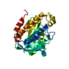
| Title | Dihydrofolate reductase from Enterococcus faecalis complexed with NADP(H)and RAB-propyl | ||||||
 Components Components | Dihydrofolate reductase | ||||||
 Keywords Keywords | OXIDOREDUCTASE / reduction of dihydrofolate to tetrahydrofolate | ||||||
| Function / homology |  Function and homology information Function and homology informationdihydrofolate metabolic process / dihydrofolate reductase / dihydrofolate reductase activity / folic acid metabolic process / tetrahydrofolate biosynthetic process / one-carbon metabolic process / NADP binding / cytosol Similarity search - Function | ||||||
| Biological species |   Enterococcus faecalis (bacteria) Enterococcus faecalis (bacteria) | ||||||
| Method |  X-RAY DIFFRACTION / MOLECULAR REPLACEMENT / Resolution: 2.3 Å X-RAY DIFFRACTION / MOLECULAR REPLACEMENT / Resolution: 2.3 Å | ||||||
 Authors Authors | Bourne, C.R. | ||||||
 Citation Citation | Journal: Biochemistry / Year: 2014 Title: The Structure and Competitive Substrate Inhibition of Dihydrofolate Reductase from Enterococcus faecalis Reveal Restrictions to Cofactor Docking. Authors: Bourne, C.R. / Wakeham, N. / Webb, N. / Nammalwar, B. / Bunce, R.A. / Berlin, K.D. / Barrow, W.W. | ||||||
| History |
|
- Structure visualization
| Structure viewer | Molecule: MolmilJmol/JSmol |
|---|
- Downloads & links
-Download
| PDBx/mmCIF format | 4m7v.cif.gz | 54.3 KB | Display | PDBx/mmCIF format |
|---|---|---|---|---|
| PDB format | pdb4m7v.ent.gz | 38 KB | Display | PDB format |
| PDBx/mmJSON format | 4m7v.json.gz | Tree view | PDBx/mmJSON format | |
| Others |  Other downloads Other downloads |
-Validation report
| Arichive directory | https://data.pdbj.org/pub/pdb/validation_reports/m7/4m7vftp://data.pdbj.org/pub/pdb/validation_reports/m7/4m7v | HTTPS FTP |
|---|
-Related structure data
| Related structure data | 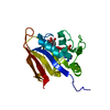 4m7uC 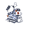 1zdrS C: citing same article ( S: Starting model for refinement |
|---|---|
| Similar structure data |

-Links
 PDBj
PDBj
- Assembly
| Deposited unit | 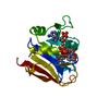
| ||||||||
|---|---|---|---|---|---|---|---|---|---|
| 1 |
| ||||||||
| Unit cell |
|
-Components
| #1: Protein | Mass: 19916.615 Da / Num. of mol.: 1 Source method: isolated from a genetically manipulated source Details: C-terminal Strep tag with cleaved thrombin site; uncleaved N-terminal TEV site Source: (gene. exp.) Enterococcus faecalis (bacteria) / Strain: 700802 / Gene: dihydrofolate reductase, EF_1577, folA / Plasmid: pPSG-IBA3 / Production host: |
|---|---|
| #2: Chemical | ChemComp-RAR /   Mass: 486.566 Da / Num. of mol.: 1 / Source method: obtained synthetically / Formula: C27H30N6O3 Mass: 486.566 Da / Num. of mol.: 1 / Source method: obtained synthetically / Formula: C27H30N6O3 |
| #3: Chemical | ChemComp-NAP /   Mass: 743.405 Da / Num. of mol.: 1 / Source method: obtained synthetically / Formula: C21H28N7O17P3 Mass: 743.405 Da / Num. of mol.: 1 / Source method: obtained synthetically / Formula: C21H28N7O17P3 |
| #4: Water | ChemComp-HOH /  Mass: 18.015 Da / Num. of mol.: 63 / Source method: isolated from a natural source / Formula: H2O Mass: 18.015 Da / Num. of mol.: 63 / Source method: isolated from a natural source / Formula: H2O |
-Experimental details
-Experiment
| Experiment | Method: X-RAY DIFFRACTION |
|---|
- Sample preparation
| Crystal | Density Matthews: 2.57 Å3/Da / Density % sol: 52.05 % |
|---|---|
| Crystal grow | Temperature: 277 K / Method: vapor diffusion, sitting drop / pH: 7 Details: 1.1 M ammonium tartrate dibasic pH 7 , VAPOR DIFFUSION, SITTING DROP, temperature 277K |
-Data collection
| Diffraction | Mean temperature: 100 K |
|---|---|
| Diffraction source | Source: ROTATING ANODE / Type: RIGAKU RUH3R / Wavelength: 1.54 Å |
| Detector | Type: RIGAKU RAXIS IV++ / Detector: IMAGE PLATE / Date: Mar 3, 2012 |
| Radiation | Protocol: SINGLE WAVELENGTH / Monochromatic (M) / Laue (L): M / Scattering type: x-ray |
| Radiation wavelength | Wavelength: 1.54 Å / Relative weight: 1 |
| Reflection | Resolution: 2.3→40.63 Å / Num. all: 9274 / Num. obs: 9220 / % possible obs: 99.4 % / Observed criterion σ(F): 0 / Redundancy: 4.2 % / Biso Wilson estimate: 35.7 Å2 / Rsym value: 0.041 / Net I/σ(I): 17.4 |
- Processing
| Software |
| ||||||||||||||||||||||||||||
|---|---|---|---|---|---|---|---|---|---|---|---|---|---|---|---|---|---|---|---|---|---|---|---|---|---|---|---|---|---|
| Refinement | Method to determine structure: MOLECULAR REPLACEMENT Starting model: SWISS-MODEL homology model based on 1ZDR Resolution: 2.3→40.628 Å / SU ML: 0.32 / σ(F): 0 / Phase error: 28.1 / Stereochemistry target values: ML
| ||||||||||||||||||||||||||||
| Solvent computation | Shrinkage radii: 1 Å / VDW probe radii: 1.2 Å / Solvent model: FLAT BULK SOLVENT MODEL | ||||||||||||||||||||||||||||
| Refinement step | Cycle: LAST / Resolution: 2.3→40.628 Å
| ||||||||||||||||||||||||||||
| Refine LS restraints |
| ||||||||||||||||||||||||||||
| LS refinement shell |
|
