 Movie
Movie Controller

[English] 日本語
 Yorodumi
- PDB-4m6g: Structure of the Mycobacterium tuberculosis peptidoglycan amidase... -
+ Open data

- Basic information
| Entry | Database: PDB / ID: 4m6g | ||||||||||||
|---|---|---|---|---|---|---|---|---|---|---|---|---|---|
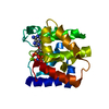
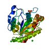
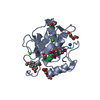
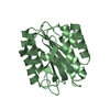
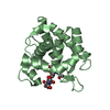
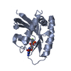
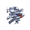
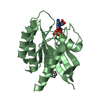
| Title | Structure of the Mycobacterium tuberculosis peptidoglycan amidase Rv3717 in complex with L-Alanine-iso-D-Glutamine reaction product | ||||||||||||
 Components Components | Peptidoglycan Amidase Rv3717 | ||||||||||||
 Keywords Keywords | HYDROLASE / Structural Genomics / NIAID / National Institute of Allergy and Infectious Diseases / TB Structural Genomics Consortium / TBSGC / Zn Binding | ||||||||||||
| Function / homology |  Function and homology information Function and homology informationN-acetylmuramoyl-L-alanine amidase / N-acetylmuramoyl-L-alanine amidase activity / FtsZ-dependent cytokinesis / peptidoglycan catabolic process / cell wall organization / cell wall macromolecule catabolic process / outer membrane-bounded periplasmic space / metal ion binding Similarity search - Function | ||||||||||||
| Biological species |   Mycobacterium tuberculosis (bacteria) Mycobacterium tuberculosis (bacteria) | ||||||||||||
| Method |  X-RAY DIFFRACTION / SYNCHROTRON / MOLECULAR REPLACEMENT / Resolution: 2.104 Å X-RAY DIFFRACTION / SYNCHROTRON / MOLECULAR REPLACEMENT / Resolution: 2.104 Å | ||||||||||||
 Authors Authors | Prigozhin, D.M. / Mavrici, D. / Huizar, J.P. / Vansell, H.J. / Alber, T. / TB Structural Genomics Consortium (TBSGC) | ||||||||||||
 Citation Citation | Journal: J.Biol.Chem. / Year: 2013 Title: Structural and Biochemical Analyses of Mycobacterium tuberculosis N-Acetylmuramyl-L-alanine Amidase Rv3717 Point to a Role in Peptidoglycan Fragment Recycling. Authors: Prigozhin, D.M. / Mavrici, D. / Huizar, J.P. / Vansell, H.J. / Alber, T. | ||||||||||||
| History |
|
- Structure visualization
| Structure viewer | Molecule: MolmilJmol/JSmol |
|---|
- Downloads & links
-Download
| PDBx/mmCIF format | 4m6g.cif.gz | 56 KB | Display | PDBx/mmCIF format |
|---|---|---|---|---|
| PDB format | pdb4m6g.ent.gz | 38.3 KB | Display | PDB format |
| PDBx/mmJSON format | 4m6g.json.gz | Tree view | PDBx/mmJSON format | |
| Others |  Other downloads Other downloads |
-Validation report
| Arichive directory | https://data.pdbj.org/pub/pdb/validation_reports/m6/4m6gftp://data.pdbj.org/pub/pdb/validation_reports/m6/4m6g | HTTPS FTP |
|---|
-Related structure data
| Related structure data |  4m6hC  4m6iC  3ne8S C: citing same article ( S: Starting model for refinement |
|---|---|
| Similar structure data |


-Links
 PDBj
PDBj- Assembly
| Deposited unit | 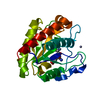
| ||||||||
|---|---|---|---|---|---|---|---|---|---|
| 1 |
| ||||||||
| Unit cell |
|
-Components
| #1: Protein | Mass: 23432.195 Da / Num. of mol.: 1 / Fragment: UNP residues 20-241 Source method: isolated from a genetically manipulated source Source: (gene. exp.) Mycobacterium tuberculosis (strain ATCC 25618 / H37Rv) (bacteria)Strain: ATCC 25618 / H37Rv / Gene: Rv3717 / Production host: References: UniProt: I6Y4D2, UniProt: O69684*PLUS, N-acetylmuramoyl-L-alanine amidase | ||||||||
|---|---|---|---|---|---|---|---|---|---|
| #2: Chemical |   Mass: 65.409 Da / Num. of mol.: 2 / Source method: obtained synthetically / Formula: Zn Mass: 65.409 Da / Num. of mol.: 2 / Source method: obtained synthetically / Formula: Zn#3: Chemical | ChemComp-ALA / |   Type: L-peptide linking / Mass: 89.093 Da / Num. of mol.: 1 / Source method: obtained synthetically / Formula: C3H7NO2 Type: L-peptide linking / Mass: 89.093 Da / Num. of mol.: 1 / Source method: obtained synthetically / Formula: C3H7NO2#4: Chemical | ChemComp-ZGL / |   Type: D-peptide linking / Mass: 146.144 Da / Num. of mol.: 1 / Source method: obtained synthetically / Formula: C5H10N2O3 Type: D-peptide linking / Mass: 146.144 Da / Num. of mol.: 1 / Source method: obtained synthetically / Formula: C5H10N2O3#5: Water | ChemComp-HOH / |  Mass: 18.015 Da / Num. of mol.: 66 / Source method: isolated from a natural source / Formula: H2O Mass: 18.015 Da / Num. of mol.: 66 / Source method: isolated from a natural source / Formula: H2OHas protein modification | Y | |
-Experimental details
-Experiment
| Experiment | Method: X-RAY DIFFRACTION / Number of used crystals: 1 |
|---|
- Sample preparation
| Crystal | Density Matthews: 2.22 Å3/Da / Density % sol: 44.67 % |
|---|---|
| Crystal grow | Temperature: 291 K / Method: vapor diffusion, hanging drop / pH: 8.5 Details: 200 mM sodium chloride, 100 mM TRIS, 25% PEG3350, pH 8.5, VAPOR DIFFUSION, HANGING DROP, temperature 291K |
-Data collection
| Diffraction | Mean temperature: 100 K |
|---|---|
| Diffraction source | Source: SYNCHROTRON / Site: ALS  / Beamline: 8.3.1 / Wavelength: 1.115869 Å / Beamline: 8.3.1 / Wavelength: 1.115869 Å |
| Detector | Type: ADSC QUANTUM 315r / Detector: CCD / Date: Apr 7, 2011 |
| Radiation | Monochromator: Double flat crystal Si(111) / Protocol: SINGLE WAVELENGTH / Monochromatic (M) / Laue (L): M / Scattering type: x-ray |
| Radiation wavelength | Wavelength: 1.115869 Å / Relative weight: 1 |
| Reflection | Resolution: 2.1→50 Å / Num. obs: 12482 / % possible obs: 97.6 % / Redundancy: 6.8 % / Biso Wilson estimate: 22.44 Å2 / Rmerge(I) obs: 0.158 / Net I/σ(I): 5.7 |
| Reflection shell | Resolution: 2.1→2.14 Å / Redundancy: 5.5 % / Mean I/σ(I) obs: 3.27 / Rsym value: 0.527 / % possible all: 87.31 |
- Processing
| Software |
| |||||||||||||||||||||||||||||||||||
|---|---|---|---|---|---|---|---|---|---|---|---|---|---|---|---|---|---|---|---|---|---|---|---|---|---|---|---|---|---|---|---|---|---|---|---|---|
| Refinement | Method to determine structure: MOLECULAR REPLACEMENT Starting model: PDB ENTRY 3NE8 Resolution: 2.104→45.065 Å / SU ML: 0.21 / σ(F): 1.35 / Phase error: 29.27 / Stereochemistry target values: ML
| |||||||||||||||||||||||||||||||||||
| Solvent computation | Shrinkage radii: 0.9 Å / VDW probe radii: 1.11 Å / Solvent model: FLAT BULK SOLVENT MODEL | |||||||||||||||||||||||||||||||||||
| Refinement step | Cycle: LAST / Resolution: 2.104→45.065 Å
| |||||||||||||||||||||||||||||||||||
| Refine LS restraints |
| |||||||||||||||||||||||||||||||||||
| LS refinement shell |
|
