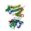
RIPK1-mediated regulated necrosis / regulation of activation-induced cell death of T cells / regulation of CD8-positive, alpha-beta cytotoxic T cell extravasation / TRIF-mediated programmed cell death / execution phase of necroptosis / regulation of T cell mediated cytotoxicity / regulation of adaptive immune response / regulation of activated T cell proliferation / Regulation of necroptotic cell death / IKK complex recruitment mediated by RIP1 ...RIPK1-mediated regulated necrosis / regulation of activation-induced cell death of T cells / regulation of CD8-positive, alpha-beta cytotoxic T cell extravasation / TRIF-mediated programmed cell death / execution phase of necroptosis / regulation of T cell mediated cytotoxicity / regulation of adaptive immune response / regulation of activated T cell proliferation / Regulation of necroptotic cell death / IKK complex recruitment mediated by RIP1 / activation of protein kinase activity / regulation of type II interferon production / programmed necrotic cell death / TRP channels / necroptotic signaling pathway / positive regulation of necroptotic process / regulation of reactive oxygen species metabolic process / non-canonical NF-kappaB signal transduction / necroptotic process / T cell homeostasis / lymph node development / spleen development / positive regulation of intrinsic apoptotic signaling pathway / thymus development / positive regulation of reactive oxygen species metabolic process / cellular response to hydrogen peroxide / T cell differentiation in thymus / regulation of apoptotic process / defense response to virus / amyloid fibril formation / protein kinase activity / non-specific serine/threonine protein kinase / protein serine kinase activity / protein serine/threonine kinase activity / apoptotic process / protein-containing complex binding / signal transduction / protein-containing complex / ATP binding / identical protein binding / nucleus / cytoplasm / cytosol Similarity search - Function
RHIM domain / RIP homotypic interaction motif / : / Transferase(Phosphotransferase) domain 1 / Transferase(Phosphotransferase); domain 1 / Serine/threonine-protein kinase, active site / Serine/Threonine protein kinases active-site signature. / Protein kinase domain / Serine/Threonine protein kinases, catalytic domain / Protein kinase, ATP binding site ...RHIM domain / RIP homotypic interaction motif / : / Transferase(Phosphotransferase) domain 1 / Transferase(Phosphotransferase); domain 1 / Serine/threonine-protein kinase, active site / Serine/Threonine protein kinases active-site signature. / Protein kinase domain / Serine/Threonine protein kinases, catalytic domain / Protein kinase, ATP binding site / Protein kinases ATP-binding region signature. / Protein kinase domain profile. / Protein kinase domain / Protein kinase-like domain superfamily / Orthogonal Bundle / Mainly Alpha Similarity search - Domain/homology
In the structure databanks used in Yorodumi, some data are registered as the other names, "COVID-19 virus" and "2019-nCoV". Here are the details of the virus and the list of structure data.
Jan 31, 2019. EMDB accession codes are about to change! (news from PDBe EMDB page)
EMDB accession codes are about to change! (news from PDBe EMDB page)
The allocation of 4 digits for EMDB accession codes will soon come to an end. Whilst these codes will remain in use, new EMDB accession codes will include an additional digit and will expand incrementally as the available range of codes is exhausted. The current 4-digit format prefixed with “EMD-” (i.e. EMD-XXXX) will advance to a 5-digit format (i.e. EMD-XXXXX), and so on. It is currently estimated that the 4-digit codes will be depleted around Spring 2019, at which point the 5-digit format will come into force.
The EM Navigator/Yorodumi systems omit the EMD- prefix.
Related info.:Q: What is EMD? / ID/Accession-code notation in Yorodumi/EM Navigator
Yorodumi is a browser for structure data from EMDB, PDB, SASBDB, etc.
This page is also the successor to EM Navigator detail page, and also detail information page/front-end page for Omokage search.
The word "yorodu" (or yorozu) is an old Japanese word meaning "ten thousand". "mi" (miru) is to see.
Related info.:EMDB / PDB / SASBDB / Comparison of 3 databanks / Yorodumi Search / Aug 31, 2016. New EM Navigator & Yorodumi / Yorodumi Papers / Jmol/JSmol / Function and homology information / Changes in new EM Navigator and Yorodumi
 Movie
Movie Controller


 Open data
 Basic information
 Components
 Keywords
 Function and homology information
 X-RAY DIFFRACTION /
  Authors
 Citation
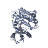
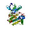
 Structure visualization
 Downloads & links
 Other downloads


 PDBj
PDBj Assembly
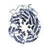
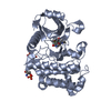
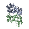

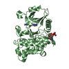
 Spodoptera frugiperda (fall armyworm)
 Mass: 18.015 Da / Num. of mol.: 106 / Source method: isolated from a natural source / Formula: H2O
 Sample preparation
 / Beamline: BL17U / Wavelength: 0.9796 Å
 Processing