 Movie
Movie Controller

[English] 日本語
 Yorodumi
- PDB-4m4w: Mechanistic implications for the bacterial primosome assembly of ... -
+ Open data

- Basic information
| Entry | Database: PDB / ID: 4m4w | ||||||
|---|---|---|---|---|---|---|---|
| Title | Mechanistic implications for the bacterial primosome assembly of the structure of a helicase-helicase loader complex | ||||||
 Components Components |
| ||||||
 Keywords Keywords | REPLICATION / primase / helicase loader / DnaB / DnaG / DnaI / DNA replication | ||||||
| Function / homology |  Function and homology information Function and homology informationDNA primase DnaG / primosome complex / DNA replication, synthesis of primer / DNA 5'-3' helicase / DNA helicase activity / DNA-directed RNA polymerase complex / Hydrolases; Acting on acid anhydrides; Acting on acid anhydrides to facilitate cellular and subcellular movement / DNA-directed RNA polymerase activity / 5'-3' DNA helicase activity / DNA replication ...DNA primase DnaG / primosome complex / DNA replication, synthesis of primer / DNA 5'-3' helicase / DNA helicase activity / DNA-directed RNA polymerase complex / Hydrolases; Acting on acid anhydrides; Acting on acid anhydrides to facilitate cellular and subcellular movement / DNA-directed RNA polymerase activity / 5'-3' DNA helicase activity / DNA replication / hydrolase activity / ATP hydrolysis activity / DNA binding / zinc ion binding / ATP binding / metal ion binding / identical protein binding / cytosol / cytoplasm Similarity search - Function | ||||||
| Biological species |   Geobacillus stearothermophilus (bacteria) Geobacillus stearothermophilus (bacteria) | ||||||
| Method |  X-RAY DIFFRACTION / SYNCHROTRON / MOLECULAR REPLACEMENT / Resolution: 6.1 Å X-RAY DIFFRACTION / SYNCHROTRON / MOLECULAR REPLACEMENT / Resolution: 6.1 Å | ||||||
 Authors Authors | Liu, B. / Eliason, W.K. / Steitz, T.A. | ||||||
 Citation Citation | Journal: Nat Commun / Year: 2013 Title: Structure of a helicase-helicase loader complex reveals insights into the mechanism of bacterial primosome assembly. Authors: Liu, B. / Eliason, W.K. / Steitz, T.A. | ||||||
| History |
|
- Structure visualization
| Structure viewer | Molecule: MolmilJmol/JSmol |
|---|
- Downloads & links
-Download
| PDBx/mmCIF format | 4m4w.cif.gz | 764.3 KB | Display | PDBx/mmCIF format |
|---|---|---|---|---|
| PDB format | pdb4m4w.ent.gz | 622.3 KB | Display | PDB format |
| PDBx/mmJSON format | 4m4w.json.gz | Tree view | PDBx/mmJSON format | |
| Others |  Other downloads Other downloads |
-Validation report
| Arichive directory | https://data.pdbj.org/pub/pdb/validation_reports/m4/4m4wftp://data.pdbj.org/pub/pdb/validation_reports/m4/4m4w | HTTPS FTP |
|---|
-Related structure data
| Similar structure data |
|---|


-Links
 PDBj
PDBj

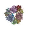
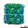
- Assembly
| Deposited unit | 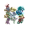
| ||||||||
|---|---|---|---|---|---|---|---|---|---|
| 1 |
| ||||||||
| Unit cell |
|
-Components
| #1: Protein | Mass: 50699.445 Da / Num. of mol.: 6 Source method: isolated from a genetically manipulated source Source: (gene. exp.) Geobacillus stearothermophilus (bacteria)Gene: dnaB / Plasmid: pET21d(+) / Production host: #2: Protein | Mass: 16796.490 Da / Num. of mol.: 3 / Fragment: Helicase Binding Domain (UNP residues 455-597) Source method: isolated from a genetically manipulated source Source: (gene. exp.) Geobacillus stearothermophilus (bacteria)Gene: dnaG / Plasmid: pET21d(+) / Production host: #3: Protein | Mass: 36991.152 Da / Num. of mol.: 6 Source method: isolated from a genetically manipulated source Source: (gene. exp.) Strain: 168 / Gene: BSU28980, dnaI, ytxA / Plasmid: pET21d(+) / Production host: Has protein modification | Y | |
|---|
-Experimental details
-Experiment
| Experiment | Method: X-RAY DIFFRACTION / Number of used crystals: 1 |
|---|
- Sample preparation
| Crystal | Density Matthews: 4.79 Å3/Da / Density % sol: 74.29 % |
|---|---|
| Crystal grow | Temperature: 290 K / Method: vapor diffusion / pH: 8 Details: 11-13% w/v PEG3350, 0.2 M lithium sulfate, 50 mM TRIS, pH 8.0-8.3, VAPOR DIFFUSION, temperature 290K |
-Data collection
| Diffraction | Mean temperature: 100 K |
|---|---|
| Diffraction source | Source: SYNCHROTRON / Site: APS  / Beamline: 24-ID-C / Wavelength: 0.97918 Å / Beamline: 24-ID-C / Wavelength: 0.97918 Å |
| Detector | Type: ADSC QUANTUM 315 / Detector: CCD / Date: Mar 11, 2010 / Details: mirrors |
| Radiation | Monochromator: Cryo-Cooled double crystal Si(111) / Protocol: SINGLE WAVELENGTH / Monochromatic (M) / Laue (L): M / Scattering type: x-ray |
| Radiation wavelength | Wavelength: 0.97918 Å / Relative weight: 1 |
| Reflection | Resolution: 6→50 Å / Num. all: 26476 / Num. obs: 26476 / % possible obs: 95.3 % / Observed criterion σ(F): 1 / Observed criterion σ(I): 1 / Redundancy: 3.1 % / Rsym value: 0.175 / Net I/σ(I): 3.7 |
| Reflection shell | Resolution: 6→6.21 Å / Redundancy: 2.5 % / Mean I/σ(I) obs: 0.5 / Num. unique all: 2472 / Rsym value: 1 / % possible all: 90.7 |
- Processing
| Software |
| |||||||||||||||||||||||||||||||||||||||||||||||||||||||||||||||||
|---|---|---|---|---|---|---|---|---|---|---|---|---|---|---|---|---|---|---|---|---|---|---|---|---|---|---|---|---|---|---|---|---|---|---|---|---|---|---|---|---|---|---|---|---|---|---|---|---|---|---|---|---|---|---|---|---|---|---|---|---|---|---|---|---|---|---|
| Refinement | Method to determine structure: MOLECULAR REPLACEMENT / Resolution: 6.1→20 Å / Cor.coef. Fo:Fc: 0.768 / Cor.coef. Fo:Fc free: 0.73 / Cross valid method: THROUGHOUT / σ(F): 0 / σ(I): 0 / ESU R Free: 3.614 / Stereochemistry target values: MAXIMUM LIKELIHOOD / Details: HYDROGENS HAVE BEEN ADDED IN THE RIDING POSITIONS
| |||||||||||||||||||||||||||||||||||||||||||||||||||||||||||||||||
| Solvent computation | Ion probe radii: 0.8 Å / Shrinkage radii: 0.8 Å / VDW probe radii: 1.4 Å / Solvent model: MASK | |||||||||||||||||||||||||||||||||||||||||||||||||||||||||||||||||
| Displacement parameters | Biso mean: 140.281 Å2
| |||||||||||||||||||||||||||||||||||||||||||||||||||||||||||||||||
| Refinement step | Cycle: LAST / Resolution: 6.1→20 Å
| |||||||||||||||||||||||||||||||||||||||||||||||||||||||||||||||||
| Refine LS restraints |
| |||||||||||||||||||||||||||||||||||||||||||||||||||||||||||||||||
| LS refinement shell | Resolution: 6.1→6.242 Å / Total num. of bins used: 20
|
