| 登録情報 | データベース: PDB / ID: 4m40
|
|---|
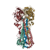
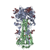
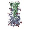
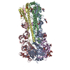
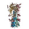
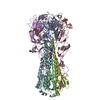
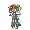
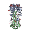
| タイトル | Crystal structure of hemagglutinin of influenza virus B/Yamanashi/166/1998 |
|---|
 要素 要素 | - Hemagglutinin HA1
- Hemagglutinin HA2
|
|---|
 キーワード キーワード | VIRAL PROTEIN / receptor binding / fusion / sialic acid |
|---|
| 機能・相同性 |  機能・相同性情報 機能・相同性情報
viral budding from plasma membrane / host cell surface receptor binding / endocytosis involved in viral entry into host cell / fusion of virus membrane with host plasma membrane / fusion of virus membrane with host endosome membrane / viral envelope / virion attachment to host cell / host cell plasma membrane / virion membrane / membrane類似検索 - 分子機能 Haemagglutinin, influenzavirus B / Hemagglutinin; Chain A, domain 2 / Hemagglutinin Chain A, Domain 2 / Hemagglutinin Ectodomain; Chain B - #10 / Hemagglutinin Ectodomain; Chain B / Hemagglutinin (Ha1 Chain); Chain: A; domain 1 / Haemagglutinin, alpha/beta domain, HA1 chain / Haemagglutinin, HA1 chain, alpha/beta domain superfamily / Haemagglutinin / Haemagglutinin, influenzavirus A/B ...Haemagglutinin, influenzavirus B / Hemagglutinin; Chain A, domain 2 / Hemagglutinin Chain A, Domain 2 / Hemagglutinin Ectodomain; Chain B - #10 / Hemagglutinin Ectodomain; Chain B / Hemagglutinin (Ha1 Chain); Chain: A; domain 1 / Haemagglutinin, alpha/beta domain, HA1 chain / Haemagglutinin, HA1 chain, alpha/beta domain superfamily / Haemagglutinin / Haemagglutinin, influenzavirus A/B / Viral capsid/haemagglutinin protein / Ribbon / Alpha-Beta Complex / Mainly Beta / Alpha Beta類似検索 - ドメイン・相同性 |
|---|
| 生物種 |  Influenza B virus (B型インフルエンザウイルス) Influenza B virus (B型インフルエンザウイルス) |
|---|
| 手法 |  X線回折 / シンクロトロン / 分子置換 / 解像度: 3.54 Å X線回折 / シンクロトロン / 分子置換 / 解像度: 3.54 Å |
|---|
 データ登録者 データ登録者 | Ni, F. / Kondrashkina, E. / Wang, Q. |
|---|
 引用 引用 | ジャーナル: Virology / 年: 2013
タイトル: Structural basis for the divergent evolution of influenza B virus hemagglutinin.
著者: Ni, F. / Kondrashkina, E. / Wang, Q. |
|---|
| 履歴 | | 登録 | 2013年8月6日 | 登録サイト: RCSB / 処理サイト: RCSB |
|---|
| 改定 1.0 | 2013年9月25日 | Provider: repository / タイプ: Initial release |
|---|
| 改定 1.1 | 2013年10月9日 | Group: Database references |
|---|
| 改定 2.0 | 2020年7月29日 | Group: Atomic model / Data collection ...Atomic model / Data collection / Database references / Derived calculations / Structure summary
カテゴリ: atom_site / chem_comp ...atom_site / chem_comp / entity / pdbx_branch_scheme / pdbx_chem_comp_identifier / pdbx_entity_branch / pdbx_entity_branch_descriptor / pdbx_entity_branch_link / pdbx_entity_branch_list / pdbx_entity_nonpoly / pdbx_nonpoly_scheme / pdbx_struct_assembly_gen / struct_asym / struct_conn / struct_ref_seq_dif / struct_site / struct_site_gen
Item: _atom_site.B_iso_or_equiv / _atom_site.Cartn_x ..._atom_site.B_iso_or_equiv / _atom_site.Cartn_x / _atom_site.Cartn_y / _atom_site.Cartn_z / _atom_site.auth_asym_id / _atom_site.auth_seq_id / _atom_site.label_asym_id / _atom_site.label_entity_id / _chem_comp.name / _chem_comp.type / _pdbx_entity_nonpoly.entity_id / _pdbx_entity_nonpoly.name / _pdbx_struct_assembly_gen.asym_id_list / _struct_conn.pdbx_dist_value / _struct_conn.pdbx_leaving_atom_flag / _struct_conn.pdbx_ptnr1_PDB_ins_code / _struct_conn.pdbx_role / _struct_conn.ptnr1_auth_asym_id / _struct_conn.ptnr1_auth_comp_id / _struct_conn.ptnr1_auth_seq_id / _struct_conn.ptnr1_label_asym_id / _struct_conn.ptnr1_label_atom_id / _struct_conn.ptnr1_label_comp_id / _struct_conn.ptnr1_label_seq_id / _struct_conn.ptnr2_auth_asym_id / _struct_conn.ptnr2_auth_comp_id / _struct_conn.ptnr2_auth_seq_id / _struct_conn.ptnr2_label_asym_id / _struct_conn.ptnr2_label_atom_id / _struct_conn.ptnr2_label_comp_id / _struct_conn.ptnr2_label_seq_id / _struct_ref_seq_dif.details
解説: Carbohydrate remediation / Provider: repository / タイプ: Remediation |
|---|
| 改定 2.1 | 2024年10月30日 | Group: Data collection / Database references / Structure summary
カテゴリ: chem_comp / chem_comp_atom ...chem_comp / chem_comp_atom / chem_comp_bond / database_2 / pdbx_entry_details / pdbx_modification_feature
Item: _chem_comp.pdbx_synonyms / _database_2.pdbx_DOI / _database_2.pdbx_database_accession |
|---|
|
|---|
 ムービー
ムービー コントローラー


 データを開く
 基本情報
基本情報 構造の表示
構造の表示 ダウンロードとリンク
 その他のダウンロード
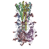


 PDBj
PDBj


 集合体
集合体

 タイプ: D-saccharide, beta linking / 分子量: 221.208 Da / 分子数: 12 / 由来タイプ: 組換発現 / 式: C8H15NO6
 試料調製
試料調製 / ビームライン: 21-ID-G / 波長: 0.97857 Å
 解析
解析