 Movie
Movie Controller

[English] 日本語
 Yorodumi
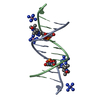
- PDB-4m3i: X-ray crystal structure of the ruthenium complex [Ru(TAP)2(dppz-{... -
+ Open data

- Basic information
| Entry | Database: PDB / ID: 4m3i | ||||||
|---|---|---|---|---|---|---|---|
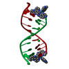
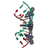
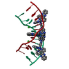
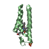
| Title | X-ray crystal structure of the ruthenium complex [Ru(TAP)2(dppz-{Me2})]2+ bound to d(CCGGTACCGG) | ||||||
 Components Components | Synthetic DNA CCGGTACCGG | ||||||
 Keywords Keywords | DNA / Intercalation / Semi-intercalation / minor-groove binder / light-switch / bending / kinking | ||||||
| Function / homology | : / Chem-RML / DNA Function and homology information Function and homology information | ||||||
| Method |  X-RAY DIFFRACTION / SYNCHROTRON / SAD / Resolution: 2.1 Å X-RAY DIFFRACTION / SYNCHROTRON / SAD / Resolution: 2.1 Å | ||||||
 Authors Authors | Niyazi, H. / Teixeira, S. / Mitchell, E. / Forsyth, T. / Cardin, C. | ||||||
 Citation Citation | Journal: To be Published Title: X-ray crystal structure of the ruthenium complex [Ru(TAP)2(dppz-{Me2})]2+ bound to d(CCGGTACCGG) Authors: Niyazi, H. / Teixeira, S. / Mitchell, E. / Forsyth, T. / Cardin, C. | ||||||
| History |
|
- Structure visualization
| Structure viewer | Molecule: MolmilJmol/JSmol |
|---|
- Downloads & links
-Download
| PDBx/mmCIF format | 4m3i.cif.gz | 19.2 KB | Display | PDBx/mmCIF format |
|---|---|---|---|---|
| PDB format | pdb4m3i.ent.gz | 12.4 KB | Display | PDB format |
| PDBx/mmJSON format | 4m3i.json.gz | Tree view | PDBx/mmJSON format | |
| Others |  Other downloads Other downloads |
-Validation report
| Arichive directory | https://data.pdbj.org/pub/pdb/validation_reports/m3/4m3iftp://data.pdbj.org/pub/pdb/validation_reports/m3/4m3i | HTTPS FTP |
|---|
-Related structure data
| Related structure data | |
|---|---|
| Similar structure data |


-Links
 PDBj
PDBj

- Assembly
| Deposited unit | 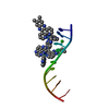
| ||||||||||||
|---|---|---|---|---|---|---|---|---|---|---|---|---|---|
| 1 | 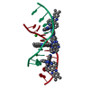
| ||||||||||||
| Unit cell |
| ||||||||||||
| Components on special symmetry positions |
|
-Components
| #1: DNA chain | Mass: 3045.992 Da / Num. of mol.: 1 / Source method: obtained synthetically / Details: Purchased Synthetic Construct | ||||
|---|---|---|---|---|---|
| #2: Chemical |   Mass: 775.785 Da / Num. of mol.: 2 / Source method: obtained synthetically / Formula: C40H26N12Ru Mass: 775.785 Da / Num. of mol.: 2 / Source method: obtained synthetically / Formula: C40H26N12Ru#3: Chemical | ChemComp-BA / |   Mass: 137.327 Da / Num. of mol.: 1 / Source method: obtained synthetically / Formula: Ba Mass: 137.327 Da / Num. of mol.: 1 / Source method: obtained synthetically / Formula: Ba#4: Water | ChemComp-HOH / |  Mass: 18.015 Da / Num. of mol.: 13 / Source method: isolated from a natural source / Formula: H2O Mass: 18.015 Da / Num. of mol.: 13 / Source method: isolated from a natural source / Formula: H2O |
-Experimental details
-Experiment
| Experiment | Method: X-RAY DIFFRACTION / Number of used crystals: 1 |
|---|
- Sample preparation
| Crystal | Density Matthews: 3.93 Å3/Da / Density % sol: 68.68 % |
|---|---|
| Crystal grow | Temperature: 293 K / Method: vapor diffusion, sitting drop / pH: 7 Details: 10ul drop containing 1ul of 4mM-[Ru(TAP)2(dppz-{Me2})]2+, 1ul of 1 mM d(CCGGTACCGG)2, and a 8ul addition containing 12mM spermine, 10% 2-methyl-2,4-pentanediol, 40mM sodium cacodylate pH 7. ...Details: 10ul drop containing 1ul of 4mM-[Ru(TAP)2(dppz-{Me2})]2+, 1ul of 1 mM d(CCGGTACCGG)2, and a 8ul addition containing 12mM spermine, 10% 2-methyl-2,4-pentanediol, 40mM sodium cacodylate pH 7.0, 80mM NaCl and 20mM BaCl2. This was equilibrated against 1ml of 35% 2-methyl-,4-pantanediol. , VAPOR DIFFUSION, SITTING DROP, temperature 293K |
-Data collection
| Diffraction | Mean temperature: 100 K |
|---|---|
| Diffraction source | Source: SYNCHROTRON / Site: ESRF  / Beamline: ID23-1 / Wavelength: 1.0332 Å / Beamline: ID23-1 / Wavelength: 1.0332 Å |
| Detector | Type: ADSC QUANTUM 315r / Detector: CCD / Date: Jul 10, 2012 |
| Radiation | Monochromator: Si(111) / Protocol: SINGLE WAVELENGTH / Monochromatic (M) / Laue (L): M / Scattering type: x-ray |
| Radiation wavelength | Wavelength: 1.0332 Å / Relative weight: 1 |
| Reflection | Resolution: 2.1→53.28 Å / Num. obs: 3114 / Observed criterion σ(I): 2 |
- Processing
| Software |
| ||||||||||||||||||||||||||||||||||||||||
|---|---|---|---|---|---|---|---|---|---|---|---|---|---|---|---|---|---|---|---|---|---|---|---|---|---|---|---|---|---|---|---|---|---|---|---|---|---|---|---|---|---|
| Refinement | Method to determine structure: SAD / Resolution: 2.1→37.67 Å / Cor.coef. Fo:Fc: 0.941 / Cor.coef. Fo:Fc free: 0.908 / SU B: 10.111 / SU ML: 0.224 / Cross valid method: THROUGHOUT / ESU R: 0.252 / ESU R Free: 0.205 / Stereochemistry target values: MAXIMUM LIKELIHOOD / Details: HYDROGENS HAVE BEEN ADDED IN THE RIDING POSITIONS
| ||||||||||||||||||||||||||||||||||||||||
| Solvent computation | Ion probe radii: 0.8 Å / Shrinkage radii: 0.8 Å / VDW probe radii: 1.2 Å / Solvent model: MASK | ||||||||||||||||||||||||||||||||||||||||
| Displacement parameters | Biso mean: 53.991 Å2
| ||||||||||||||||||||||||||||||||||||||||
| Refinement step | Cycle: LAST / Resolution: 2.1→37.67 Å
| ||||||||||||||||||||||||||||||||||||||||
| Refine LS restraints |
| ||||||||||||||||||||||||||||||||||||||||
| LS refinement shell | Resolution: 2.1→2.155 Å / Total num. of bins used: 20
|
