 Movie
Movie Controller

[English] 日本語
 Yorodumi
- PDB-3qrn: X-ray crystal structure of the ruthenium complex [Ru(tap)2(dppz)]... -
+ Open data

- Basic information
| Entry | Database: PDB / ID: 3qrn | ||||||||||||||||||
|---|---|---|---|---|---|---|---|---|---|---|---|---|---|---|---|---|---|---|---|
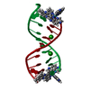
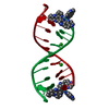
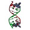
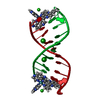
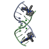
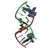
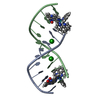
| Title | X-ray crystal structure of the ruthenium complex [Ru(tap)2(dppz)]2+ bound to d(TCGGCGCCGA)at high resolution | ||||||||||||||||||
 Components Components | 5'-D(* Keywords KeywordsDNA / intercalation / semi-intercalation / B-DNA | Function / homology | : / Ru(tap)2(dppz) complex / DNA |  Function and homology information Function and homology informationMethod |  X-RAY DIFFRACTION / SYNCHROTRON / MOLECULAR REPLACEMENT / Resolution: 1.1 Å X-RAY DIFFRACTION / SYNCHROTRON / MOLECULAR REPLACEMENT / Resolution: 1.1 Å  Authors AuthorsCardin, C.J. / Hall, J.P. |  CitationJournal: Proc.Natl.Acad.Sci.USA / Year: 2011 CitationJournal: Proc.Natl.Acad.Sci.USA / Year: 2011Title: Structure determination of an intercalating ruthenium dipyridophenazine complex which kinks DNA by semiintercalation of a tetraazaphenanthrene ligand. Authors: Hall, J.P. / O'Sullivan, K. / Naseer, A. / Smith, J.A. / Kelly, J.M. / Cardin, C.J. History |
|
- Structure visualization
| Structure viewer | Molecule: MolmilJmol/JSmol |
|---|
- Downloads & links
-Download
| PDBx/mmCIF format | 3qrn.cif.gz | 27.7 KB | Display | PDBx/mmCIF format |
|---|---|---|---|---|
| PDB format | pdb3qrn.ent.gz | 18.7 KB | Display | PDB format |
| PDBx/mmJSON format | 3qrn.json.gz | Tree view | PDBx/mmJSON format | |
| Others |  Other downloads Other downloads |
-Validation report
| Arichive directory | https://data.pdbj.org/pub/pdb/validation_reports/qr/3qrnftp://data.pdbj.org/pub/pdb/validation_reports/qr/3qrn | HTTPS FTP |
|---|
-Related structure data
| Related structure data |  3gomC  3qf8SC S: Starting model for refinement C: citing same article ( |
|---|---|
| Similar structure data |

-Links
 PDBj
PDBj

- Assembly
| Deposited unit | 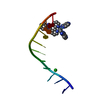
| ||||||||
|---|---|---|---|---|---|---|---|---|---|
| 1 | 
| ||||||||
| Unit cell |
|
-Components
| #1: DNA chain | Mass: 3045.992 Da / Num. of mol.: 1 / Source method: obtained synthetically |
|---|---|
| #2: Chemical | ChemComp-RKL /   Mass: 747.732 Da / Num. of mol.: 1 / Source method: obtained synthetically / Formula: C38H22N12Ru Mass: 747.732 Da / Num. of mol.: 1 / Source method: obtained synthetically / Formula: C38H22N12Ru |
| #3: Chemical | ChemComp-BA /   Mass: 137.327 Da / Num. of mol.: 1 / Source method: obtained synthetically / Formula: Ba Mass: 137.327 Da / Num. of mol.: 1 / Source method: obtained synthetically / Formula: Ba |
| #4: Water | ChemComp-HOH /  Mass: 18.015 Da / Num. of mol.: 64 / Source method: isolated from a natural source / Formula: H2O Mass: 18.015 Da / Num. of mol.: 64 / Source method: isolated from a natural source / Formula: H2O |
-Experimental details
-Experiment
| Experiment | Method: X-RAY DIFFRACTION / Number of used crystals: 1 |
|---|
- Sample preparation
| Crystal | Density Matthews: 2.93 Å3/Da / Density % sol: 58.03 % |
|---|---|
| Crystal grow | Temperature: 276 K / Method: vapor diffusion, sitting drop / pH: 7 Details: 10%(v/v) 2-methyl-2,4-pentanediol, 40mM sodium cacodylate, 12mM spermine tetra-HCl, 80mM sodium chloride, 20mM barium chloride, 1mM DNA d(TCGGCGCCGA), 1mM lambda-[Ru(tap)2dppz)Cl2, pH 7, ...Details: 10%(v/v) 2-methyl-2,4-pentanediol, 40mM sodium cacodylate, 12mM spermine tetra-HCl, 80mM sodium chloride, 20mM barium chloride, 1mM DNA d(TCGGCGCCGA), 1mM lambda-[Ru(tap)2dppz)Cl2, pH 7, VAPOR DIFFUSION, SITTING DROP, temperature 276K |
-Data collection
| Diffraction | Mean temperature: 100 K |
|---|---|
| Diffraction source | Source: SYNCHROTRON / Site: Diamond  / Beamline: I02 / Wavelength: 0.9795 Å / Beamline: I02 / Wavelength: 0.9795 Å |
| Detector | Type: ADSC QUANTUM 315r / Detector: CCD / Date: Feb 16, 2011 |
| Radiation | Monochromator: Si(111) double crystal / Protocol: SINGLE WAVELENGTH / Monochromatic (M) / Laue (L): M / Scattering type: x-ray |
| Radiation wavelength | Wavelength: 0.9795 Å / Relative weight: 1 |
| Reflection | Resolution: 1.1→21.16 Å / Num. all: 15135 / Num. obs: 14312 / % possible obs: 88.14 % / Observed criterion σ(I): 3 / Rmerge(I) obs: 0.066 |
| Reflection shell | Resolution: 1.1→1.16 Å / Redundancy: 16 % / Rmerge(I) obs: 0.452 / Mean I/σ(I) obs: 6.1 / Num. unique all: 2160 / % possible all: 100 |
- Processing
| Software |
| |||||||||||||||||||||||||
|---|---|---|---|---|---|---|---|---|---|---|---|---|---|---|---|---|---|---|---|---|---|---|---|---|---|---|
| Refinement | Method to determine structure: MOLECULAR REPLACEMENT Starting model: 3QF8 Resolution: 1.1→18.93 Å / Cor.coef. Fo:Fc: 0.982 / Cor.coef. Fo:Fc free: 0.978 / SU ML: 0.011 / Cross valid method: THROUGHOUT / ESU R Free: 0.021 / Stereochemistry target values: MAXIMUM LIKELIHOOD
| |||||||||||||||||||||||||
| Solvent computation | Ion probe radii: 0.8 Å / Shrinkage radii: 0.8 Å / VDW probe radii: 1.4 Å / Solvent model: MASK | |||||||||||||||||||||||||
| Refinement step | Cycle: LAST / Resolution: 1.1→18.93 Å
| |||||||||||||||||||||||||
| LS refinement shell | Resolution: 1.102→1.131 Å / Total num. of bins used: 20
|
