 Movie
Movie Controller

[English] 日本語
 Yorodumi
- PDB-4m2p: Crystal structure of a non-myristoylated C39D recoverin mutant wi... -
+ Open data

- Basic information
| Entry | Database: PDB / ID: 4m2p | ||||||
|---|---|---|---|---|---|---|---|
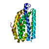
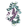
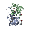
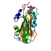
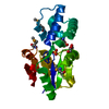
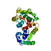
| Title | Crystal structure of a non-myristoylated C39D recoverin mutant with one calcium ion bound to EF-hand 3 | ||||||
 Components Components | Recoverin | ||||||
 Keywords Keywords | METAL BINDING PROTEIN / CALCIUM BINDING PROTEIN / EF HAND / NEURONAL CALCIUM SENSING (NCS) FAMILY PROTEIN / INHIBITS RHODOPSIN KINASE / RHODOPSIN KINASE / RETINA | ||||||
| Function / homology |  Function and homology information Function and homology informationInactivation, recovery and regulation of the phototransduction cascade / regulation of calcium ion transport / phototransduction / photoreceptor outer segment / regulation of signal transduction / photoreceptor inner segment / visual perception / perikaryon / calcium ion binding / membrane / cytosol Similarity search - Function | ||||||
| Biological species |  | ||||||
| Method |  X-RAY DIFFRACTION / SYNCHROTRON / MOLECULAR REPLACEMENT / molecular replacement / Resolution: 1.45 Å X-RAY DIFFRACTION / SYNCHROTRON / MOLECULAR REPLACEMENT / molecular replacement / Resolution: 1.45 Å | ||||||
 Authors Authors | Prem Kumar, R. / Ranaghan, M.J. / Oprian, D.D. | ||||||
 Citation Citation | Journal: J.Biol.Chem. / Year: 2013 Title: A Highly Conserved Cysteine of Neuronal Calcium-sensing Proteins Controls Cooperative Binding of Ca2+ to Recoverin. Authors: Ranaghan, M.J. / Kumar, R.P. / Chakrabarti, K.S. / Buosi, V. / Kern, D. / Oprian, D.D. | ||||||
| History |
|
- Structure visualization
| Structure viewer | Molecule: MolmilJmol/JSmol |
|---|
- Downloads & links
-Download
| PDBx/mmCIF format | 4m2p.cif.gz | 58.3 KB | Display | PDBx/mmCIF format |
|---|---|---|---|---|
| PDB format | pdb4m2p.ent.gz | 41.2 KB | Display | PDB format |
| PDBx/mmJSON format | 4m2p.json.gz | Tree view | PDBx/mmJSON format | |
| Others |  Other downloads Other downloads |
-Validation report
| Summary document | 4m2p_validation.pdf.gz | 428.2 KB | Display | wwPDB validaton report |
|---|---|---|---|---|
| Full document | 4m2p_full_validation.pdf.gz | 433.6 KB | Display | |
| Data in XML | 4m2p_validation.xml.gz | 10.9 KB | Display | |
| Data in CIF | 4m2p_validation.cif.gz | 15.1 KB | Display | |
| Arichive directory | https://data.pdbj.org/pub/pdb/validation_reports/m2/4m2pftp://data.pdbj.org/pub/pdb/validation_reports/m2/4m2p | HTTPS FTP |
-Related structure data
| Related structure data | 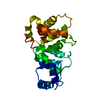 4m2oC 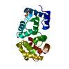 4m2qC 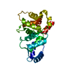 4mlwC 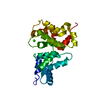 1omrS S: Starting model for refinement C: citing same article ( |
|---|---|
| Similar structure data |


-Links
 PDBj
PDBj


- Assembly
| Deposited unit | 
| ||||||||
|---|---|---|---|---|---|---|---|---|---|
| 1 |
| ||||||||
| Unit cell |
|
-Components
| #1: Protein | Mass: 23247.152 Da / Num. of mol.: 1 / Mutation: C39D Source method: isolated from a genetically manipulated source Source: (gene. exp.)  |
|---|---|
| #2: Chemical | ChemComp-CA /   Mass: 40.078 Da / Num. of mol.: 1 / Source method: obtained synthetically / Formula: Ca Mass: 40.078 Da / Num. of mol.: 1 / Source method: obtained synthetically / Formula: Ca |
| #3: Water | ChemComp-HOH /  Mass: 18.015 Da / Num. of mol.: 142 / Source method: isolated from a natural source / Formula: H2O Mass: 18.015 Da / Num. of mol.: 142 / Source method: isolated from a natural source / Formula: H2O |
-Experimental details
-Experiment
| Experiment | Method: X-RAY DIFFRACTION / Number of used crystals: 1 |
|---|
- Sample preparation
| Crystal | Density Matthews: 2.27 Å3/Da / Density % sol: 45.89 % |
|---|---|
| Crystal grow | Temperature: 296 K / Method: vapor diffusion, hanging drop / pH: 7 Details: 2.4 M SODIUM MALONATE, pH 7.0, VAPOR DIFFUSION, HANGING DROP, temperature 296K |
-Data collection
| Diffraction | Mean temperature: 100 K |
|---|---|
| Diffraction source | Source: SYNCHROTRON / Site: ALS  / Beamline: 8.2.1 / Wavelength: 1 / Beamline: 8.2.1 / Wavelength: 1 |
| Detector | Type: ADSC QUANTUM 315r / Detector: CCD / Date: May 15, 2013 / Details: MIRRORS |
| Radiation | Monochromator: DOUBLE CRYSTAL SI(111) / Protocol: SINGLE WAVELENGTH / Monochromatic (M) / Laue (L): M / Scattering type: x-ray |
| Radiation wavelength | Wavelength: 1 Å / Relative weight: 1 |
| Reflection | Resolution: 1.45→32 Å / Num. obs: 36942 / % possible obs: 100 % / Observed criterion σ(I): 2 / Redundancy: 4.1 % / Biso Wilson estimate: 66.4 Å2 / Rmerge(I) obs: 0.095 / Net I/σ(I): 9.6 |
| Reflection shell | Resolution: 1.45→1.53 Å / Redundancy: 3.6 % / Rmerge(I) obs: 0.421 / Mean I/σ(I) obs: 2.6 / % possible all: 100 |
-Phasing
| Phasing | Method: molecular replacement |
|---|
- Processing
| Software |
| ||||||||||||||||||||||||||||||||||||||||||||||||||||||||||||||||||||||||||||||||||||||||||||||||||
|---|---|---|---|---|---|---|---|---|---|---|---|---|---|---|---|---|---|---|---|---|---|---|---|---|---|---|---|---|---|---|---|---|---|---|---|---|---|---|---|---|---|---|---|---|---|---|---|---|---|---|---|---|---|---|---|---|---|---|---|---|---|---|---|---|---|---|---|---|---|---|---|---|---|---|---|---|---|---|---|---|---|---|---|---|---|---|---|---|---|---|---|---|---|---|---|---|---|---|---|
| Refinement | Method to determine structure: MOLECULAR REPLACEMENT Starting model: pdb entry 1OMR Resolution: 1.45→29.8 Å / Occupancy max: 1 / Occupancy min: 0.55 / SU ML: 0.16 / σ(F): 1.35 / Phase error: 22.28 / Stereochemistry target values: ML
| ||||||||||||||||||||||||||||||||||||||||||||||||||||||||||||||||||||||||||||||||||||||||||||||||||
| Solvent computation | Shrinkage radii: 0.9 Å / VDW probe radii: 1.11 Å / Solvent model: FLAT BULK SOLVENT MODEL | ||||||||||||||||||||||||||||||||||||||||||||||||||||||||||||||||||||||||||||||||||||||||||||||||||
| Displacement parameters | Biso mean: 62.4 Å2 | ||||||||||||||||||||||||||||||||||||||||||||||||||||||||||||||||||||||||||||||||||||||||||||||||||
| Refinement step | Cycle: LAST / Resolution: 1.45→29.8 Å
| ||||||||||||||||||||||||||||||||||||||||||||||||||||||||||||||||||||||||||||||||||||||||||||||||||
| Refine LS restraints |
| ||||||||||||||||||||||||||||||||||||||||||||||||||||||||||||||||||||||||||||||||||||||||||||||||||
| LS refinement shell |
|
