 Movie
Movie Controller

[English] 日本語
 Yorodumi
- PDB-4lxj: Saccharomyces cerevisiae lanosterol 14-alpha demethylase with lan... -
+ Open data

- Basic information
| Entry | Database: PDB / ID: 4lxj | ||||||
|---|---|---|---|---|---|---|---|
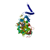
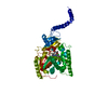
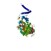
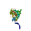
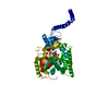
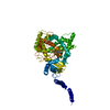
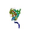
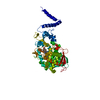
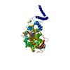
| Title | Saccharomyces cerevisiae lanosterol 14-alpha demethylase with lanosterol bound | ||||||
 Components Components | Lanosterol 14-alpha demethylase | ||||||
 Keywords Keywords | OXIDOREDUCTASE / Cytochrome p450 | ||||||
| Function / homology |  Function and homology information Function and homology informationEndogenous sterols / Cholesterol biosynthesis via desmosterol (Bloch pathway) / sterol 14alpha-demethylase / ergosterol biosynthetic process / sterol 14-demethylase activity / oxidoreductase activity / iron ion binding / heme binding / endoplasmic reticulum membrane / endoplasmic reticulum Similarity search - Function | ||||||
| Biological species |  | ||||||
| Method |  X-RAY DIFFRACTION / SYNCHROTRON / MOLECULAR REPLACEMENT / Resolution: 1.9 Å X-RAY DIFFRACTION / SYNCHROTRON / MOLECULAR REPLACEMENT / Resolution: 1.9 Å | ||||||
 Authors Authors | Monk, B.C. / Tomasiak, T.M. / Keniya, M.V. / Huschmann, F.U. / Tyndall, J.D.A. / O'Connell III, J.D. / Cannon, R.D. / McDonald, J. / Rodriguez, A. / Finer-Moore, J. / Stroud, R.M. | ||||||
 Citation Citation | Journal: Proc.Natl.Acad.Sci.USA / Year: 2014 Title: Architecture of a single membrane spanning cytochrome P450 suggests constraints that orient the catalytic domain relative to a bilayer. Authors: Monk, B.C. / Tomasiak, T.M. / Keniya, M.V. / Huschmann, F.U. / Tyndall, J.D. / O'Connell, J.D. / Cannon, R.D. / McDonald, J.G. / Rodriguez, A. / Finer-Moore, J.S. / Stroud, R.M. | ||||||
| History |
|
- Structure visualization
| Structure viewer | Molecule: MolmilJmol/JSmol |
|---|
- Downloads & links
-Download
| PDBx/mmCIF format | 4lxj.cif.gz | 126.4 KB | Display | PDBx/mmCIF format |
|---|---|---|---|---|
| PDB format | pdb4lxj.ent.gz | 96.4 KB | Display | PDB format |
| PDBx/mmJSON format | 4lxj.json.gz | Tree view | PDBx/mmJSON format | |
| Others |  Other downloads Other downloads |
-Validation report
| Arichive directory | https://data.pdbj.org/pub/pdb/validation_reports/lx/4lxjftp://data.pdbj.org/pub/pdb/validation_reports/lx/4lxj | HTTPS FTP |
|---|
-Related structure data
| Related structure data |  5eqbC 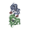 3ld6S S: Starting model for refinement C: citing same article ( |
|---|---|
| Similar structure data |

-Links
 PDBj
PDBj


- Assembly
| Deposited unit | 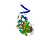
| ||||||||
|---|---|---|---|---|---|---|---|---|---|
| 1 |
| ||||||||
| Unit cell |
|
-Components
| #1: Protein | Mass: 61470.766 Da / Num. of mol.: 1 Source method: isolated from a genetically manipulated source Source: (gene. exp.) Gene: ERG11, CYP51, YHR007C / Production host: |
|---|---|
| #2: Chemical | ChemComp-HEM /   Mass: 616.487 Da / Num. of mol.: 1 / Source method: obtained synthetically / Formula: C34H32FeN4O4 Mass: 616.487 Da / Num. of mol.: 1 / Source method: obtained synthetically / Formula: C34H32FeN4O4 |
| #3: Chemical | ChemComp-LAN /   Mass: 426.717 Da / Num. of mol.: 1 / Source method: obtained synthetically / Formula: C30H50O Mass: 426.717 Da / Num. of mol.: 1 / Source method: obtained synthetically / Formula: C30H50O |
| #4: Chemical | ChemComp-OXY /   Mass: 31.999 Da / Num. of mol.: 1 / Source method: obtained synthetically / Formula: O2 Mass: 31.999 Da / Num. of mol.: 1 / Source method: obtained synthetically / Formula: O2 |
| #5: Water | ChemComp-HOH /  Mass: 18.015 Da / Num. of mol.: 182 / Source method: isolated from a natural source / Formula: H2O Mass: 18.015 Da / Num. of mol.: 182 / Source method: isolated from a natural source / Formula: H2O |
-Experimental details
-Experiment
| Experiment | Method: X-RAY DIFFRACTION / Number of used crystals: 1 |
|---|
- Sample preparation
| Crystal | Density Matthews: 3.38 Å3/Da / Density % sol: 63.6 % |
|---|---|
| Crystal grow | Temperature: 291 K / Method: vapor diffusion / pH: 9 Details: 49% PEG 400, 100mM Glycine pH 9.0, VAPOR DIFFUSION, temperature 291K |
-Data collection
| Diffraction | Mean temperature: 100 K |
|---|---|
| Diffraction source | Source: SYNCHROTRON / Site: ALS  / Beamline: 8.3.1 / Beamline: 8.3.1 |
| Detector | Type: ADSC QUANTUM 315r / Detector: CCD |
| Radiation | Monochromator: Khozu double flat crystal / Protocol: SINGLE WAVELENGTH / Monochromatic (M) / Laue (L): M / Scattering type: x-ray |
| Radiation wavelength | Relative weight: 1 |
| Reflection | Resolution: 1.9→50 Å / Num. all: 64791 / Num. obs: 62523 / % possible obs: 96.5 % / Observed criterion σ(F): -3 / Observed criterion σ(I): -1 / Redundancy: 3.5 % / Rmerge(I) obs: 0.045 |
| Reflection shell | Resolution: 1.9→1.93 Å / Redundancy: 2.2 % / Rsym value: 0.531 / % possible all: 72.8 |
- Processing
| Software |
| |||||||||||||||||||||||||||||||||||||||||||||||||||||||||||||||||||||||||||||||||||||||||||||||||||||||||||||||||||||||||||||||||||||||||||||||||||||||||||||||||
|---|---|---|---|---|---|---|---|---|---|---|---|---|---|---|---|---|---|---|---|---|---|---|---|---|---|---|---|---|---|---|---|---|---|---|---|---|---|---|---|---|---|---|---|---|---|---|---|---|---|---|---|---|---|---|---|---|---|---|---|---|---|---|---|---|---|---|---|---|---|---|---|---|---|---|---|---|---|---|---|---|---|---|---|---|---|---|---|---|---|---|---|---|---|---|---|---|---|---|---|---|---|---|---|---|---|---|---|---|---|---|---|---|---|---|---|---|---|---|---|---|---|---|---|---|---|---|---|---|---|---|---|---|---|---|---|---|---|---|---|---|---|---|---|---|---|---|---|---|---|---|---|---|---|---|---|---|---|---|---|---|---|---|
| Refinement | Method to determine structure: MOLECULAR REPLACEMENT Starting model: PDB entry 3LD6 Resolution: 1.9→29.327 Å / SU ML: 0.2 / σ(F): 1.38 / Phase error: 26.16 / Stereochemistry target values: ML Details: VISUAL INSPECTION OF THE ELECTRON DENSITY MAPS AND A HIGH REAL SPACE R-FACTOR INDICATED THE NEED FOR CONSIDERABLE CARE IN PLACEMENT OF THE LIGAND IN THE LANOSTEROL CO-STRUCTURE. MINIMALLY ...Details: VISUAL INSPECTION OF THE ELECTRON DENSITY MAPS AND A HIGH REAL SPACE R-FACTOR INDICATED THE NEED FOR CONSIDERABLE CARE IN PLACEMENT OF THE LIGAND IN THE LANOSTEROL CO-STRUCTURE. MINIMALLY BIASED FO-FC ELECTRON DENSITY OMIT MAPS, CALCULATED WITH LANOSTEROL OMITTED TO CORRECTLY PLACE THE LANOSTEROL, RESULTED IN A REASONABLE CORRELATION COEFFICIENT (0.813).
| |||||||||||||||||||||||||||||||||||||||||||||||||||||||||||||||||||||||||||||||||||||||||||||||||||||||||||||||||||||||||||||||||||||||||||||||||||||||||||||||||
| Solvent computation | Shrinkage radii: 0.9 Å / VDW probe radii: 1.11 Å / Solvent model: FLAT BULK SOLVENT MODEL | |||||||||||||||||||||||||||||||||||||||||||||||||||||||||||||||||||||||||||||||||||||||||||||||||||||||||||||||||||||||||||||||||||||||||||||||||||||||||||||||||
| Refinement step | Cycle: LAST / Resolution: 1.9→29.327 Å
| |||||||||||||||||||||||||||||||||||||||||||||||||||||||||||||||||||||||||||||||||||||||||||||||||||||||||||||||||||||||||||||||||||||||||||||||||||||||||||||||||
| Refine LS restraints |
| |||||||||||||||||||||||||||||||||||||||||||||||||||||||||||||||||||||||||||||||||||||||||||||||||||||||||||||||||||||||||||||||||||||||||||||||||||||||||||||||||
| LS refinement shell |
|
