Mass: 18.015 Da / Num. of mol.: 165 / Source method: isolated from a natural source / Formula: H2O
Has protein modification
Y
-
Experimental details
-
Experiment
Experiment
Method: X-RAY DIFFRACTION / Number of used crystals: 1
-
Sample preparation
Crystal
Density Matthews: 2.46 Å3/Da / Density % sol: 49.98 %
Crystal grow
Temperature: 295 K / pH: 7.5 Details: Protein solutions at a concentration of 20mg/ml were mixed with the reservoir solutions of 35-44% polyethylene glycol 8000 in 100mM HEPES pH 7.5 buffer containing 100mM NaBr in the 1:1, 2:1 ...Details: Protein solutions at a concentration of 20mg/ml were mixed with the reservoir solutions of 35-44% polyethylene glycol 8000 in 100mM HEPES pH 7.5 buffer containing 100mM NaBr in the 1:1, 2:1 and 3:1 volume ratios and vapor diffused against the reservoir solutions. Plate-shaped crystals appeared in about a week and continued to grow for a few more weeks., VAPOR DIFFUSION, SITTING DROP, temperature 295.K
Resolution: 1.9→50 Å / Cor.coef. Fo:Fc: 0.946 / Cor.coef. Fo:Fc free: 0.918 / SU B: 4.273 / SU ML: 0.126 / Cross valid method: THROUGHOUT / ESU R: 0.174 / ESU R Free: 0.165 / Stereochemistry target values: MAXIMUM LIKELIHOOD / Details: HYDROGENS HAVE BEEN ADDED IN THE RIDING POSITIONS
Rfactor
Num. reflection
% reflection
Selection details
Rfree
0.268
1332
5 %
RANDOM
Rwork
0.219
-
-
-
obs
0.222
25467
98.3 %
-
all
-
27277
-
-
Solvent computation
Ion probe radii: 0.8 Å / Shrinkage radii: 0.8 Å / VDW probe radii: 1.4 Å / Solvent model: MASK
 Movie
Movie Controller

 Yorodumi
 Open data
 Basic information
 Components
 Keywords
 Function and homology information
 X-RAY DIFFRACTION /
  Authors
 Citation
 Structure visualization
 Downloads & links
 Other downloads

 PDBj
PDBj


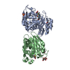
 Assembly
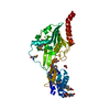


 Mass: 238.305 Da / Num. of mol.: 1 / Source method: obtained synthetically / Formula: C8H18N2O4S / Comment: pH buffer*YM
 Mass: 92.094 Da / Num. of mol.: 3 / Source method: obtained synthetically / Formula: C3H8O3
 Mass: 282.461 Da / Num. of mol.: 1 / Source method: obtained synthetically / Formula: C18H34O2
 Mass: 18.015 Da / Num. of mol.: 165 / Source method: isolated from a natural source / Formula: H2O
 Sample preparation
 / Beamline: 19-ID / Wavelength: 0.979368, 0.979484, 0.9716932
 Processing