 Movie
Movie Controller

[English] 日本語
 Yorodumi
- PDB-4lsw: Crystallization and Structural Analysis of 2-Hydroxyacid Dehydrog... -
+ Open data

- Basic information
| Entry | Database: PDB / ID: 4lsw | ||||||
|---|---|---|---|---|---|---|---|
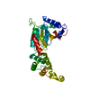
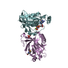
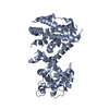
| Title | Crystallization and Structural Analysis of 2-Hydroxyacid Dehydrogenase from Ketogulonicigenium vulgare Y25 | ||||||
 Components Components | D-2-hydroxyacid dehydrogensase protein | ||||||
 Keywords Keywords | HYDROLASE / hydrogenase | ||||||
| Function / homology |  Function and homology information Function and homology informationoxidoreductase activity, acting on the CH-OH group of donors, NAD or NADP as acceptor / NAD binding Similarity search - Function | ||||||
| Biological species |  Ketogulonicigenium vulgare (bacteria) Ketogulonicigenium vulgare (bacteria) | ||||||
| Method |  X-RAY DIFFRACTION / SYNCHROTRON / MOLECULAR REPLACEMENT / Resolution: 1.64 Å X-RAY DIFFRACTION / SYNCHROTRON / MOLECULAR REPLACEMENT / Resolution: 1.64 Å | ||||||
 Authors Authors | Han, X. / Liu, X. | ||||||
 Citation Citation | Journal: Biotechnol.Lett. / Year: 2014 Title: Crystallization and structural analysis of 2-hydroxyacid dehydrogenase from Ketogulonicigenium vulgare. Authors: Han, X. / Xiong, X. / Hu, X. / Li, M. / Zhang, W. / Liu, X. | ||||||
| History |
|
- Structure visualization
| Structure viewer | Molecule: MolmilJmol/JSmol |
|---|
- Downloads & links
-Download
| PDBx/mmCIF format | 4lsw.cif.gz | 82.8 KB | Display | PDBx/mmCIF format |
|---|---|---|---|---|
| PDB format | pdb4lsw.ent.gz | 61.3 KB | Display | PDB format |
| PDBx/mmJSON format | 4lsw.json.gz | Tree view | PDBx/mmJSON format | |
| Others |  Other downloads Other downloads |
-Validation report
| Arichive directory | https://data.pdbj.org/pub/pdb/validation_reports/ls/4lswftp://data.pdbj.org/pub/pdb/validation_reports/ls/4lsw | HTTPS FTP |
|---|
-Related structure data
| Related structure data |  3ba1S S: Starting model for refinement |
|---|---|
| Similar structure data |


-Links
 PDBj
PDBj
- Assembly
| Deposited unit | 
| ||||||||||||
|---|---|---|---|---|---|---|---|---|---|---|---|---|---|
| 1 |
| ||||||||||||
| 2 | 
| ||||||||||||
| Unit cell |
| ||||||||||||
| Components on special symmetry positions |
|
-Components
| #1: Protein | Mass: 34394.133 Da / Num. of mol.: 1 / Source method: isolated from a natural source / Source: (natural) Ketogulonicigenium vulgare (bacteria) / Strain: Y25 / References: UniProt: E3F052 |
|---|---|
| #2: Water | ChemComp-HOH /  Mass: 18.015 Da / Num. of mol.: 430 / Source method: isolated from a natural source / Formula: H2O Mass: 18.015 Da / Num. of mol.: 430 / Source method: isolated from a natural source / Formula: H2O |
-Experimental details
-Experiment
| Experiment | Method: X-RAY DIFFRACTION / Number of used crystals: 6 |
|---|
- Sample preparation
| Crystal | Density Matthews: 2.65 Å3/Da / Density % sol: 53.57 % Description: THE ENTRY CONTAINS FRIEDEL PAIRS IN I_PLUS/MINUS COLUMNS |
|---|---|
| Crystal grow | Temperature: 298 K / Method: vapor diffusion, sitting drop / pH: 7 Details: 23% PEG 3350, 0.2M MgCl, 0.1M pH 7.0 HEPES, VAPOR DIFFUSION, SITTING DROP, temperature 298K |
-Data collection
| Diffraction | Mean temperature: 100 K | ||||||||||||||||||
|---|---|---|---|---|---|---|---|---|---|---|---|---|---|---|---|---|---|---|---|
| Diffraction source | Source: SYNCHROTRON / Site: BSRF  / Beamline: 1W2B / Wavelength: 0.9792 Å / Beamline: 1W2B / Wavelength: 0.9792 Å | ||||||||||||||||||
| Detector | Type: MAR scanner 345 mm plate / Detector: IMAGE PLATE / Date: Oct 31, 2012 | ||||||||||||||||||
| Radiation | Protocol: SINGLE WAVELENGTH / Monochromatic (M) / Laue (L): M / Scattering type: x-ray | ||||||||||||||||||
| Radiation wavelength | Wavelength: 0.9792 Å / Relative weight: 1 | ||||||||||||||||||
| Reflection | Resolution: 1.64→50 Å / Num. obs: 38014 / % possible obs: 85 % / Observed criterion σ(F): 2 / Observed criterion σ(I): 1 | ||||||||||||||||||
| Reflection shell |
|
- Processing
| Software |
| ||||||||||||||||||||||||||||||||||||||||||||||||||||||||||||||||||||||||||||||||||||||||||
|---|---|---|---|---|---|---|---|---|---|---|---|---|---|---|---|---|---|---|---|---|---|---|---|---|---|---|---|---|---|---|---|---|---|---|---|---|---|---|---|---|---|---|---|---|---|---|---|---|---|---|---|---|---|---|---|---|---|---|---|---|---|---|---|---|---|---|---|---|---|---|---|---|---|---|---|---|---|---|---|---|---|---|---|---|---|---|---|---|---|---|---|
| Refinement | Method to determine structure: MOLECULAR REPLACEMENT Starting model: 3BA1 Resolution: 1.64→50 Å / SU ML: 0.23 / σ(F): 2 / Phase error: 21.82 / Stereochemistry target values: ML Details: THE ENTRY CONTAINS FRIEDEL PAIRS IN I_PLUS/MINUS COLUMNS
| ||||||||||||||||||||||||||||||||||||||||||||||||||||||||||||||||||||||||||||||||||||||||||
| Solvent computation | Shrinkage radii: 0.47 Å / VDW probe radii: 0.8 Å / Solvent model: FLAT BULK SOLVENT MODEL / Bsol: 33.584 Å2 / ksol: 0.435 e/Å3 | ||||||||||||||||||||||||||||||||||||||||||||||||||||||||||||||||||||||||||||||||||||||||||
| Displacement parameters |
| ||||||||||||||||||||||||||||||||||||||||||||||||||||||||||||||||||||||||||||||||||||||||||
| Refinement step | Cycle: LAST / Resolution: 1.64→50 Å
| ||||||||||||||||||||||||||||||||||||||||||||||||||||||||||||||||||||||||||||||||||||||||||
| Refine LS restraints |
| ||||||||||||||||||||||||||||||||||||||||||||||||||||||||||||||||||||||||||||||||||||||||||
| LS refinement shell | Refine-ID: X-RAY DIFFRACTION / Total num. of bins used: 14
|
