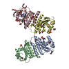
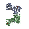
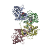
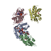
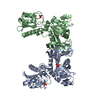
- PDB-2hxv: Crystal structure of a diaminohydroxyphosphoribosylaminopyrimidin... -
+
Open data
ID or keywords:
Loading...
-
Basic information
Entry
Database: PDB / ID: 2hxv
Title
Crystal structure of a diaminohydroxyphosphoribosylaminopyrimidine deaminase/ 5-amino-6-(5-phosphoribosylamino)uracil reductase (tm1828) from thermotoga maritima at 1.80 A resolution
BIOMOLECULE: 1 THIS ENTRY CONTAINS THE CRYSTALLOGRAPHIC ASYMMETRIC UNIT WHICH CONSISTS OF 1 CHAIN(S) ...BIOMOLECULE: 1 THIS ENTRY CONTAINS THE CRYSTALLOGRAPHIC ASYMMETRIC UNIT WHICH CONSISTS OF 1 CHAIN(S). SEE REMARK 350 FOR INFORMATION ON GENERATING THE BIOLOGICAL MOLECULE(S). SIZE EXCLUSION CHROMATOGRAPHY SUPPORTS THE ASSIGNMENT OF A DIMER AS THE BIOLOGICALLY SIGNIFICANT OLIGIMERIZATION STATE.
Remark 999
SEQUENCE THE CONSTRUCT WAS EXPRESSED WITH A PURIFICATION TAG MGSDKIHHHHHH.
Monochromator: Single crystal Si(111) bent monochromator (horizontal focusing) Protocol: MAD / Monochromatic (M) / Laue (L): M / Scattering type: x-ray
Radiation wavelength
ID
Wavelength (Å)
Relative weight
1
1.000001
1
2
0.97917
1
3
0.91837
1
4
0.978913
1
Reflection
Resolution: 1.8→28.341 Å / Num. obs: 37367 / % possible obs: 99.7 % / Redundancy: 4.945 % / Biso Wilson estimate: 30.526 Å2 / Rmerge(I) obs: 0.072 / Net I/σ(I): 14.96
Reflection shell
Diffraction-ID: 1
Resolution (Å)
Highest resolution (Å)
Redundancy (%)
Rmerge(I) obs
Mean I/σ(I) obs
Num. measured obs
Num. unique all
% possible all
1.8-1.86
3.711
0.719
1.71
22207
5938
97.7
1.86-1.94
0.506
2.6
26451
7031
92
1.94-2.03
0.375
3.5
25888
6857
95
2.03-2.13
0.319
4.8
28217
6423
96.7
2.13-2.27
0.23
7
35038
7275
97.3
2.27-2.44
0.179
9.6
37652
6816
98.7
2.44-2.69
0.13
13.4
42415
7168
99.1
2.69-3.07
0.08
20.6
40986
6937
99.4
3.07
0.043
35
42101
7176
99.7
-
Phasing
Phasing
Method: MAD
-
Processing
Software
Name
Version
Classification
NB
MolProbity
3beta29
modelbuilding
SHELX
phasing
REFMAC
5.2.0005
refinement
XSCALE
datascaling
PDB_EXTRACT
2
dataextraction
XDS
datareduction
SHARP
phasing
Refinement
Method to determine structure: MAD / Resolution: 1.8→28.341 Å / Cor.coef. Fo:Fc: 0.967 / Cor.coef. Fo:Fc free: 0.955 / SU B: 4.664 / SU ML: 0.073 / TLS residual ADP flag: LIKELY RESIDUAL / Cross valid method: THROUGHOUT / σ(F): 0 / ESU R: 0.105 / ESU R Free: 0.104 Stereochemistry target values: MAXIMUM LIKELIHOOD WITH PHASES Details: 1. HYDROGENS HAVE BEEN ADDED IN THE RIDING POSITIONS. 2. A MET-INHIBITION PROTOCOL WAS USED FOR SELENOMETHIONINE INCORPORATION DURING PROTEIN EXPRESSION. THE OCCUPANCY OF THE SE ATOMS IN THE ...Details: 1. HYDROGENS HAVE BEEN ADDED IN THE RIDING POSITIONS. 2. A MET-INHIBITION PROTOCOL WAS USED FOR SELENOMETHIONINE INCORPORATION DURING PROTEIN EXPRESSION. THE OCCUPANCY OF THE SE ATOMS IN THE MSE RESIDUES WAS REDUCED TO 0.75 TO ACCOUNT FOR THE REDUCED SCATTERING POWER DUE TO PARTIAL S-MET INCORPORATION. 3. ATOM RECORD CONTAINS RESIDUAL B FACTORS ONLY. 4. NADPH WAS MODELED BASED ON DENSITY AND PROPOSED FUNCTION. 5. ZN METAL IDENTIFICATION SUPPORTED BY EXCITATION AND FLOURESENCE SCAN ANALYSIS, IN ADDITION TO GEOMETRY AND HOMOLOGS. 6.GLYCEROL AND CL WERE MODELED BASED ON CRYSTALLIZATION CONDITIONS. 7. RESIDUES 80-81 WERE MODELED IN VERY WEAK DENSITY. 8. UNKNOWN DENSITIES NEAR LYS 14 AND LYS 304 WERE NOT MODELED.
Rfactor
Num. reflection
% reflection
Selection details
Rfree
0.195
1877
5 %
RANDOM
Rwork
0.16
-
-
-
obs
0.162
37365
99.84 %
-
Solvent computation
Ion probe radii: 0.8 Å / Shrinkage radii: 0.8 Å / VDW probe radii: 1.2 Å / Solvent model: MASK
Displacement parameters
Biso mean: 28.964 Å2
Baniso -1
Baniso -2
Baniso -3
1-
0.46 Å2
0 Å2
0 Å2
2-
-
0.46 Å2
0 Å2
3-
-
-
-0.92 Å2
Refinement step
Cycle: LAST / Resolution: 1.8→28.341 Å
Protein
Nucleic acid
Ligand
Solvent
Total
Num. atoms
2697
0
98
205
3000
Refine LS restraints
Refine-ID
Type
Dev ideal
Dev ideal target
Number
X-RAY DIFFRACTION
r_bond_refined_d
0.017
0.022
2873
X-RAY DIFFRACTION
r_bond_other_d
0.001
0.02
2639
X-RAY DIFFRACTION
r_angle_refined_deg
1.69
1.994
3895
X-RAY DIFFRACTION
r_angle_other_deg
0.999
3
6124
X-RAY DIFFRACTION
r_dihedral_angle_1_deg
5.695
5
358
X-RAY DIFFRACTION
r_dihedral_angle_2_deg
31.676
23.388
121
X-RAY DIFFRACTION
r_dihedral_angle_3_deg
12.044
15
466
X-RAY DIFFRACTION
r_dihedral_angle_4_deg
14.474
15
19
X-RAY DIFFRACTION
r_chiral_restr
0.099
0.2
434
X-RAY DIFFRACTION
r_gen_planes_refined
0.008
0.02
3139
X-RAY DIFFRACTION
r_gen_planes_other
0.001
0.02
576
X-RAY DIFFRACTION
r_nbd_refined
0.209
0.2
544
X-RAY DIFFRACTION
r_nbd_other
0.19
0.2
2786
X-RAY DIFFRACTION
r_nbtor_refined
0.188
0.2
1445
X-RAY DIFFRACTION
r_nbtor_other
0.087
0.2
1766
X-RAY DIFFRACTION
r_xyhbond_nbd_refined
0.19
0.2
172
X-RAY DIFFRACTION
r_symmetry_vdw_refined
0.148
0.2
15
X-RAY DIFFRACTION
r_symmetry_vdw_other
0.257
0.2
80
X-RAY DIFFRACTION
r_symmetry_hbond_refined
0.177
0.2
19
X-RAY DIFFRACTION
r_mcbond_it
2.415
3
1796
X-RAY DIFFRACTION
r_mcbond_other
0.641
3
716
X-RAY DIFFRACTION
r_mcangle_it
3.353
5
2840
X-RAY DIFFRACTION
r_scbond_it
5.473
8
1197
X-RAY DIFFRACTION
r_scangle_it
7.869
11
1050
LS refinement shell
Resolution: 1.8→1.847 Å / Total num. of bins used: 20
Rfactor
Num. reflection
% reflection
Rfree
0.304
127
-
Rwork
0.217
2536
-
obs
-
2663
98.7 %
Refinement TLS params.
Method: refined / Refine-ID: X-RAY DIFFRACTION
ID
L11 (°2)
L12 (°2)
L13 (°2)
L22 (°2)
L23 (°2)
L33 (°2)
S11 (Å °)
S12 (Å °)
S13 (Å °)
S21 (Å °)
S22 (Å °)
S23 (Å °)
S31 (Å °)
S32 (Å °)
S33 (Å °)
T11 (Å2)
T12 (Å2)
T13 (Å2)
T22 (Å2)
T23 (Å2)
T33 (Å2)
Origin x (Å)
Origin y (Å)
Origin z (Å)
1
0.8457
1.0195
-0.0024
3.0063
-0.1763
0.182
0.0325
0.0215
0.0484
0.0132
-0.0256
0.1208
-0.0016
-0.0261
-0.0069
-0.058
-0.0005
-0.0073
-0.0883
-0.0178
-0.0689
26.527
-3.033
22.838
2
0.3364
-0.0552
0.4115
0.1889
-0.337
1.4062
-0.0292
0.0137
0.0151
0.0058
-0.0261
-0.0041
-0.0604
0.1278
0.0554
-0.0985
-0.022
0.0021
-0.0686
-0.0175
-0.0605
37.27
27.888
15.955
Refinement TLS group
Refine-ID: X-RAY DIFFRACTION / Selection: ALL / Auth asym-ID: A / Label asym-ID: A
ID
Refine TLS-ID
Auth seq-ID
Label seq-ID
1
1
-3 - 146
9 - 158
2
2
147 - 345
159 - 357
+
About Yorodumi
-
News
-
Feb 9, 2022. New format data for meta-information of EMDB entries
New format data for meta-information of EMDB entries
Version 3 of the EMDB header file is now the official format.
The previous official version 1.9 will be removed from the archive.
In the structure databanks used in Yorodumi, some data are registered as the other names, "COVID-19 virus" and "2019-nCoV". Here are the details of the virus and the list of structure data.
Jan 31, 2019. EMDB accession codes are about to change! (news from PDBe EMDB page)
EMDB accession codes are about to change! (news from PDBe EMDB page)
The allocation of 4 digits for EMDB accession codes will soon come to an end. Whilst these codes will remain in use, new EMDB accession codes will include an additional digit and will expand incrementally as the available range of codes is exhausted. The current 4-digit format prefixed with “EMD-” (i.e. EMD-XXXX) will advance to a 5-digit format (i.e. EMD-XXXXX), and so on. It is currently estimated that the 4-digit codes will be depleted around Spring 2019, at which point the 5-digit format will come into force.
The EM Navigator/Yorodumi systems omit the EMD- prefix.
Related info.:Q: What is EMD? / ID/Accession-code notation in Yorodumi/EM Navigator
Yorodumi is a browser for structure data from EMDB, PDB, SASBDB, etc.
This page is also the successor to EM Navigator detail page, and also detail information page/front-end page for Omokage search.
The word "yorodu" (or yorozu) is an old Japanese word meaning "ten thousand". "mi" (miru) is to see.
Related info.:EMDB / PDB / SASBDB / Comparison of 3 databanks / Yorodumi Search / Aug 31, 2016. New EM Navigator & Yorodumi / Yorodumi Papers / Jmol/JSmol / Function and homology information / Changes in new EM Navigator and Yorodumi
 Movie
Movie Controller

 Yorodumi
 Open data
 Basic information
 Components
 Keywords
 Function and homology information
 Thermotoga maritima (bacteria)
 X-RAY DIFFRACTION /
  Authors
 Citation
 Structure visualization
 Downloads & links
 Other downloads
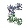


 PDBj
PDBj

 Assembly
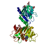


 Mass: 65.409 Da / Num. of mol.: 1 / Source method: obtained synthetically / Formula: Zn
 Mass: 35.453 Da / Num. of mol.: 1 / Source method: obtained synthetically / Formula: Cl
 Mass: 745.421 Da / Num. of mol.: 1 / Source method: obtained synthetically / Formula: C21H30N7O17P3
 Mass: 92.094 Da / Num. of mol.: 8 / Source method: obtained synthetically / Formula: C3H8O3
 Sample preparation
 Processing