 Movie
Movie Controller

[English] 日本語
 Yorodumi
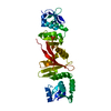
- PDB-4lr4: Crystal structure of a putative secreted protein (EUBREC_3654) fr... -
+ Open data

- Basic information
| Entry | Database: PDB / ID: 4lr4 | ||||||
|---|---|---|---|---|---|---|---|
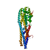
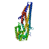
| Title | Crystal structure of a putative secreted protein (EUBREC_3654) from Eubacterium rectale at 2.43 A resolution | ||||||
 Components Components | hypothetical protein | ||||||
 Keywords Keywords | STRUCTURAL GENOMICS / UNKNOWN FUNCTION / an orphan / two domains protein / N-terminus has a sandwich 10 strands in 2 sheets and jelly-roll fold / C -terminus has galactose-binding domain-like fold / Joint Center for Structural Genomics / JCSG / Protein Structure Initiative / PSI-BIOLOGY | ||||||
| Function / homology |  Function and homology information Function and homology informationJelly Rolls - #1420 / Jelly Rolls - #1430 / : / : / Carbohydrate binding module-like / Domain of unknown function (DUF6947) / Jelly Rolls / Sandwich / Mainly Beta Similarity search - Domain/homology | ||||||
| Biological species |  Eubacterium rectale (bacteria) Eubacterium rectale (bacteria) | ||||||
| Method |  X-RAY DIFFRACTION / SYNCHROTRON / MAD / Resolution: 2.43 Å X-RAY DIFFRACTION / SYNCHROTRON / MAD / Resolution: 2.43 Å | ||||||
 Authors Authors | Joint Center for Structural Genomics (JCSG) | ||||||
 Citation Citation | Journal: To be published Title: Crystal structure of a hypothetical protein (EUBREC_3654) from Eubacterium rectale at 2.43 A resolution Authors: Joint Center for Structural Genomics (JCSG) | ||||||
| History |
|
- Structure visualization
| Structure viewer | Molecule: MolmilJmol/JSmol |
|---|
- Downloads & links
-Download
| PDBx/mmCIF format | 4lr4.cif.gz | 567.6 KB | Display | PDBx/mmCIF format |
|---|---|---|---|---|
| PDB format | pdb4lr4.ent.gz | 470.2 KB | Display | PDB format |
| PDBx/mmJSON format | 4lr4.json.gz | Tree view | PDBx/mmJSON format | |
| Others |  Other downloads Other downloads |
-Validation report
| Summary document | 4lr4_validation.pdf.gz | 495.2 KB | Display | wwPDB validaton report |
|---|---|---|---|---|
| Full document | 4lr4_full_validation.pdf.gz | 505.6 KB | Display | |
| Data in XML | 4lr4_validation.xml.gz | 56.2 KB | Display | |
| Data in CIF | 4lr4_validation.cif.gz | 80.3 KB | Display | |
| Arichive directory | https://data.pdbj.org/pub/pdb/validation_reports/lr/4lr4ftp://data.pdbj.org/pub/pdb/validation_reports/lr/4lr4 | HTTPS FTP |
-Related structure data
| Similar structure data | |
|---|---|
| Other databases |


-Links
 PDBj
PDBj- Assembly
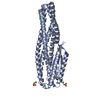
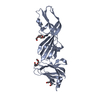
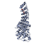
| Deposited unit | 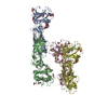
| ||||||||
|---|---|---|---|---|---|---|---|---|---|
| 1 | 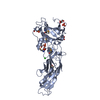
| ||||||||
| 2 | 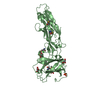
| ||||||||
| 3 | 
| ||||||||
| 4 | 
| ||||||||
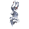
| Unit cell |
|
-Components
| #1: Protein | Mass: 41218.746 Da / Num. of mol.: 4 Source method: isolated from a genetically manipulated source Source: (gene. exp.) Eubacterium rectale (bacteria) / Strain: ATCC 33656 / VPI 0990 / Gene: EUBREC_3654, RER070207000171 / Plasmid: SpeedET / Production host: #2: Chemical | ChemComp-GOL /   Mass: 92.094 Da / Num. of mol.: 17 / Source method: obtained synthetically / Formula: C3H8O3 Mass: 92.094 Da / Num. of mol.: 17 / Source method: obtained synthetically / Formula: C3H8O3#3: Chemical | ChemComp-PO4 /   Mass: 94.971 Da / Num. of mol.: 10 / Source method: obtained synthetically / Formula: PO4 Mass: 94.971 Da / Num. of mol.: 10 / Source method: obtained synthetically / Formula: PO4#4: Chemical | ChemComp-CL /   Mass: 35.453 Da / Num. of mol.: 9 / Source method: obtained synthetically / Formula: Cl Mass: 35.453 Da / Num. of mol.: 9 / Source method: obtained synthetically / Formula: Cl#5: Water | ChemComp-HOH / |  Mass: 18.015 Da / Num. of mol.: 683 / Source method: isolated from a natural source / Formula: H2O Mass: 18.015 Da / Num. of mol.: 683 / Source method: isolated from a natural source / Formula: H2OHas protein modification | Y | Sequence details | THE CONSTRUCT (26-394) WAS EXPRESSED WITH A PURIFICATION TAG MGSDKIHHHHHHENLYFQG. THE TAG WAS ...THE CONSTRUCT (26-394) WAS EXPRESSED WITH A PURIFICATI | |
|---|
-Experimental details
-Experiment
| Experiment | Method: X-RAY DIFFRACTION / Number of used crystals: 1 |
|---|
- Sample preparation
| Crystal | Density Matthews: 3.65 Å3/Da / Density % sol: 66.28 % |
|---|---|
| Crystal grow | Temperature: 277 K / Method: vapor diffusion, sitting drop / pH: 7.83 Details: 1.5M ammonium dihydrogen phosphate, 20.0% Glycerol, 0.01M magnesium chloride, 0.1M TRIS pH 7.83, NANODROP, VAPOR DIFFUSION, SITTING DROP, temperature 277K |
-Data collection
| Diffraction | Mean temperature: 100 K | |||||||||||||||||||||||||||||||||||||||||||||||||||||||||||||||||||||||||||||
|---|---|---|---|---|---|---|---|---|---|---|---|---|---|---|---|---|---|---|---|---|---|---|---|---|---|---|---|---|---|---|---|---|---|---|---|---|---|---|---|---|---|---|---|---|---|---|---|---|---|---|---|---|---|---|---|---|---|---|---|---|---|---|---|---|---|---|---|---|---|---|---|---|---|---|---|---|---|---|
| Diffraction source | Source: SYNCHROTRON / Site: SSRL  / Beamline: BL11-1 / Wavelength: 0.91837, 0.97864, 0.97805 / Beamline: BL11-1 / Wavelength: 0.91837, 0.97864, 0.97805 | |||||||||||||||||||||||||||||||||||||||||||||||||||||||||||||||||||||||||||||
| Detector | Type: DECTRIS PILATUS 6M / Detector: PIXEL / Date: May 2, 2013 Details: Flat mirror (vertical focusing); single crystal Si(111) bent monochromator (horizontal focusing) | |||||||||||||||||||||||||||||||||||||||||||||||||||||||||||||||||||||||||||||
| Radiation | Monochromator: single crystal Si(111) bent / Protocol: MAD / Monochromatic (M) / Laue (L): M / Scattering type: x-ray | |||||||||||||||||||||||||||||||||||||||||||||||||||||||||||||||||||||||||||||
| Radiation wavelength |
| |||||||||||||||||||||||||||||||||||||||||||||||||||||||||||||||||||||||||||||
| Reflection | Resolution: 2.43→48.963 Å / Num. obs: 87733 / % possible obs: 98.1 % / Observed criterion σ(I): -3 / Biso Wilson estimate: 52.762 Å2 / Rmerge(I) obs: 0.102 / Net I/σ(I): 9.58 | |||||||||||||||||||||||||||||||||||||||||||||||||||||||||||||||||||||||||||||
| Reflection shell | Diffraction-ID: 1
|
-Phasing
| Phasing | Method: MAD |
|---|
- Processing
| Software |
| |||||||||||||||||||||||||||||||||||||||||||||||||||||||||||||||||||||||||||||||||||||||||||||||||||||||||||||||||||||||||||||
|---|---|---|---|---|---|---|---|---|---|---|---|---|---|---|---|---|---|---|---|---|---|---|---|---|---|---|---|---|---|---|---|---|---|---|---|---|---|---|---|---|---|---|---|---|---|---|---|---|---|---|---|---|---|---|---|---|---|---|---|---|---|---|---|---|---|---|---|---|---|---|---|---|---|---|---|---|---|---|---|---|---|---|---|---|---|---|---|---|---|---|---|---|---|---|---|---|---|---|---|---|---|---|---|---|---|---|---|---|---|---|---|---|---|---|---|---|---|---|---|---|---|---|---|---|---|---|
| Refinement | Method to determine structure: MAD / Resolution: 2.43→48.963 Å / Cor.coef. Fo:Fc: 0.9314 / Cor.coef. Fo:Fc free: 0.9191 / Occupancy max: 1 / Occupancy min: 0.5 / Cross valid method: THROUGHOUT / σ(F): 0 Details: 1. A MET-INHIBITION PROTOCOL WAS USED FOR SELENOMETHIONINE INCORPORATION DURING PROTEIN EXPRESSION. THE OCCUPANCY OF THE SE ATOMS IN THE MSE RESIDUES WAS REDUCED TO 0.75 FOR THE REDUCED ...Details: 1. A MET-INHIBITION PROTOCOL WAS USED FOR SELENOMETHIONINE INCORPORATION DURING PROTEIN EXPRESSION. THE OCCUPANCY OF THE SE ATOMS IN THE MSE RESIDUES WAS REDUCED TO 0.75 FOR THE REDUCED SCATTERING POWER DUE TO PARTIAL S-MET INCORPORATION. 2. ATOM RECORD CONTAINS SUM OF TLS AND RESIDUAL B FACTORS. ANISOU RECORD CONTAINS SUM OF TLS AND RESIDUAL U FACTORS. 3. THE MAD PHASES WERE USED AS RESTRAINTS DURING REFINEMENT. 4. PHOSPHATE (PO4), GLYCEROL (GOL), AND CHLORIDE (CL) MODELED WERE PRESENT IN PROTEIN/CYRO CONDITIONS. 5.NCS RESTRAINTS WERE IMPOSED BY AUTOBUSTER'S LSSR PROCEDURE (-AUTONCS). 6. RAMACHANDRAN OUTLIERS (A,D/378, A-D/181) ARE SUPPORTED BY DENSITY.
| |||||||||||||||||||||||||||||||||||||||||||||||||||||||||||||||||||||||||||||||||||||||||||||||||||||||||||||||||||||||||||||
| Displacement parameters | Biso max: 179.94 Å2 / Biso mean: 52.857 Å2 / Biso min: 20.29 Å2
| |||||||||||||||||||||||||||||||||||||||||||||||||||||||||||||||||||||||||||||||||||||||||||||||||||||||||||||||||||||||||||||
| Refine analyze | Luzzati coordinate error obs: 0.348 Å | |||||||||||||||||||||||||||||||||||||||||||||||||||||||||||||||||||||||||||||||||||||||||||||||||||||||||||||||||||||||||||||
| Refinement step | Cycle: LAST / Resolution: 2.43→48.963 Å
| |||||||||||||||||||||||||||||||||||||||||||||||||||||||||||||||||||||||||||||||||||||||||||||||||||||||||||||||||||||||||||||
| Refine LS restraints |
| |||||||||||||||||||||||||||||||||||||||||||||||||||||||||||||||||||||||||||||||||||||||||||||||||||||||||||||||||||||||||||||
| LS refinement shell | Resolution: 2.43→2.49 Å / Total num. of bins used: 20
| |||||||||||||||||||||||||||||||||||||||||||||||||||||||||||||||||||||||||||||||||||||||||||||||||||||||||||||||||||||||||||||
| Refinement TLS params. | Method: refined / Refine-ID: X-RAY DIFFRACTION
| |||||||||||||||||||||||||||||||||||||||||||||||||||||||||||||||||||||||||||||||||||||||||||||||||||||||||||||||||||||||||||||
| Refinement TLS group |
|
