 Movie
Movie Controller

[English] 日本語
 Yorodumi
- PDB-4lqw: Crystal structure of HIV-1 capsid N-terminal domain in complex wi... -
+ Open data

- Basic information
| Entry | Database: PDB / ID: 4lqw | ||||||
|---|---|---|---|---|---|---|---|
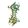
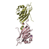
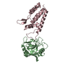
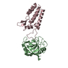
| Title | Crystal structure of HIV-1 capsid N-terminal domain in complex with NUP358 cyclophilin | ||||||
 Components Components |
| ||||||
 Keywords Keywords | ISOMERASE / cyclophilin / capsid | ||||||
| Function / homology |  Function and homology information Function and homology informationcytoplasmic periphery of the nuclear pore complex / SUMO ligase complex / SUMO ligase activity / annulate lamellae / nuclear pore cytoplasmic filaments / Nuclear Pore Complex (NPC) Disassembly / Regulation of Glucokinase by Glucokinase Regulatory Protein / Defective TPR may confer susceptibility towards thyroid papillary carcinoma (TPC) / nuclear inclusion body / Transport of Ribonucleoproteins into the Host Nucleus ...cytoplasmic periphery of the nuclear pore complex / SUMO ligase complex / SUMO ligase activity / annulate lamellae / nuclear pore cytoplasmic filaments / Nuclear Pore Complex (NPC) Disassembly / Regulation of Glucokinase by Glucokinase Regulatory Protein / Defective TPR may confer susceptibility towards thyroid papillary carcinoma (TPC) / nuclear inclusion body / Transport of Ribonucleoproteins into the Host Nucleus / nuclear pore nuclear basket / Transport of the SLBP independent Mature mRNA / Transport of the SLBP Dependant Mature mRNA / SUMOylation of SUMOylation proteins / NS1 Mediated Effects on Host Pathways / Transport of Mature mRNA Derived from an Intronless Transcript / Rev-mediated nuclear export of HIV RNA / nuclear export / Nuclear import of Rev protein / SUMOylation of RNA binding proteins / NEP/NS2 Interacts with the Cellular Export Machinery / Transferases; Acyltransferases; Aminoacyltransferases / SUMO transferase activity / Transport of Mature mRNA derived from an Intron-Containing Transcript / tRNA processing in the nucleus / kinase activator activity / centrosome localization / nucleocytoplasmic transport / regulation of gluconeogenesis / Viral Messenger RNA Synthesis / NLS-bearing protein import into nucleus / SUMOylation of ubiquitinylation proteins / Vpr-mediated nuclear import of PICs / SUMOylation of DNA replication proteins / Regulation of HSF1-mediated heat shock response / protein sumoylation / nuclear pore / mRNA transport / SUMOylation of DNA damage response and repair proteins / Amplification of signal from unattached kinetochores via a MAD2 inhibitory signal / Mitotic Prometaphase / EML4 and NUDC in mitotic spindle formation / intracellular glucose homeostasis / response to amphetamine / Resolution of Sister Chromatid Cohesion / SUMOylation of chromatin organization proteins / HCMV Late Events / HIV-1 retropepsin / symbiont-mediated activation of host apoptosis / retroviral ribonuclease H / exoribonuclease H / exoribonuclease H activity / Transcriptional regulation by small RNAs / DNA integration / RHO GTPases Activate Formins / viral genome integration into host DNA / establishment of integrated proviral latency / RNA-directed DNA polymerase / RNA stem-loop binding / viral penetration into host nucleus / host multivesicular body / RNA-directed DNA polymerase activity / RNA-DNA hybrid ribonuclease activity / ISG15 antiviral mechanism / small GTPase binding / Transferases; Transferring phosphorus-containing groups; Nucleotidyltransferases / HCMV Early Events / Signaling by ALK fusions and activated point mutants / Separation of Sister Chromatids / nuclear envelope / host cell / viral nucleocapsid / protein folding / nuclear membrane / DNA recombination / snRNP Assembly / DNA-directed DNA polymerase / aspartic-type endopeptidase activity / Hydrolases; Acting on ester bonds / DNA-directed DNA polymerase activity / protein-macromolecule adaptor activity / symbiont-mediated suppression of host gene expression / viral translational frameshifting / symbiont entry into host cell / lipid binding / host cell nucleus / host cell plasma membrane / protein-containing complex binding / SARS-CoV-2 activates/modulates innate and adaptive immune responses / virion membrane / structural molecule activity / negative regulation of transcription by RNA polymerase II / proteolysis / DNA binding / RNA binding / zinc ion binding / nucleoplasm / membrane / nucleus / cytosol Similarity search - Function | ||||||
| Biological species |  Homo sapiens (human) Homo sapiens (human)  Human immunodeficiency virus type 1 Human immunodeficiency virus type 1 | ||||||
| Method |  X-RAY DIFFRACTION / SYNCHROTRON / MOLECULAR REPLACEMENT / Resolution: 1.95 Å X-RAY DIFFRACTION / SYNCHROTRON / MOLECULAR REPLACEMENT / Resolution: 1.95 Å | ||||||
 Authors Authors | Price, A.J. / James, L.C. | ||||||
 Citation Citation | Journal: Retrovirology / Year: 2013 Title: HIV-1 capsid undergoes coupled binding and isomerization by the nuclear pore protein NUP358. Authors: Bichel, K. / Price, A.J. / Schaller, T. / Towers, G.J. / Freund, S.M. / James, L.C. | ||||||
| History |
|
- Structure visualization
| Structure viewer | Molecule: MolmilJmol/JSmol |
|---|
- Downloads & links
-Download
| PDBx/mmCIF format | 4lqw.cif.gz | 248.6 KB | Display | PDBx/mmCIF format |
|---|---|---|---|---|
| PDB format | pdb4lqw.ent.gz | 202.3 KB | Display | PDB format |
| PDBx/mmJSON format | 4lqw.json.gz | Tree view | PDBx/mmJSON format | |
| Others |  Other downloads Other downloads |
-Validation report
| Arichive directory | https://data.pdbj.org/pub/pdb/validation_reports/lq/4lqwftp://data.pdbj.org/pub/pdb/validation_reports/lq/4lqw | HTTPS FTP |
|---|
-Related structure data
| Related structure data | 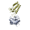 1ak4S S: Starting model for refinement |
|---|---|
| Similar structure data |


-Links
 PDBj
PDBj


- Assembly
| Deposited unit | 
| ||||||||
|---|---|---|---|---|---|---|---|---|---|
| 1 | 
| ||||||||
| 2 | 
| ||||||||
| Unit cell |
|
-Components
| #1: Protein | Mass: 19713.320 Da / Num. of mol.: 2 / Fragment: UNP Residues 3057-3224 Source method: isolated from a genetically manipulated source Source: (gene. exp.) Homo sapiens (human) / Gene: RANBP2, NUP358 / Production host:  References: UniProt: P49792, Ligases; Forming carbon-nitrogen bonds; Acid-amino-acid ligases (peptide synthases), peptidylprolyl isomerase #2: Protein | Mass: 16204.573 Da / Num. of mol.: 2 / Fragment: UNP Residues 133-278 Source method: isolated from a genetically manipulated source Source: (gene. exp.) Human immunodeficiency virus type 1 / Gene: gag-pol / Production host: #3: Water | ChemComp-HOH / |  Mass: 18.015 Da / Num. of mol.: 372 / Source method: isolated from a natural source / Formula: H2O Mass: 18.015 Da / Num. of mol.: 372 / Source method: isolated from a natural source / Formula: H2O |
|---|
-Experimental details
-Experiment
| Experiment | Method: X-RAY DIFFRACTION / Number of used crystals: 1 |
|---|
- Sample preparation
| Crystal | Density Matthews: 2.3 Å3/Da / Density % sol: 46.6 % |
|---|---|
| Crystal grow | Temperature: 290 K / Method: vapor diffusion, sitting drop / pH: 7.5 Details: 23 % v/v PEG 4000, 23 % glycerol, 8.5 % isopropanol, 85 mM HEPES pH 7.5, 20 mM spermine tetrahydrochloride, 100 mM glycine, VAPOR DIFFUSION, SITTING DROP, temperature 290K |
-Data collection
| Diffraction | Mean temperature: 100 K |
|---|---|
| Diffraction source | Source: SYNCHROTRON / Site: ESRF  / Beamline: ID14-1 / Wavelength: 0.934 Å / Beamline: ID14-1 / Wavelength: 0.934 Å |
| Detector | Type: ADSC QUANTUM 210 / Detector: CCD / Date: May 1, 2009 |
| Radiation | Monochromator: DIAMOND(111) + Ge(220) / Protocol: SINGLE WAVELENGTH / Monochromatic (M) / Laue (L): M / Scattering type: x-ray |
| Radiation wavelength | Wavelength: 0.934 Å / Relative weight: 1 |
| Reflection | Resolution: 1.953→33.04 Å / Num. all: 48326 / Num. obs: 48326 / % possible obs: 94.5 % / Observed criterion σ(F): 0 / Observed criterion σ(I): 0 |
| Reflection shell | Resolution: 1.953→2.004 Å / % possible all: 88.43 |
- Processing
| Software |
| ||||||||||||||||||||||||||||||||||||||||||||||||||||||||||||||||||||||||||||||||||||||||||||||||||||||||||||||||||||||||||||||||||||||||||||||||||||||||||||||||||||||||||||||||||||||
|---|---|---|---|---|---|---|---|---|---|---|---|---|---|---|---|---|---|---|---|---|---|---|---|---|---|---|---|---|---|---|---|---|---|---|---|---|---|---|---|---|---|---|---|---|---|---|---|---|---|---|---|---|---|---|---|---|---|---|---|---|---|---|---|---|---|---|---|---|---|---|---|---|---|---|---|---|---|---|---|---|---|---|---|---|---|---|---|---|---|---|---|---|---|---|---|---|---|---|---|---|---|---|---|---|---|---|---|---|---|---|---|---|---|---|---|---|---|---|---|---|---|---|---|---|---|---|---|---|---|---|---|---|---|---|---|---|---|---|---|---|---|---|---|---|---|---|---|---|---|---|---|---|---|---|---|---|---|---|---|---|---|---|---|---|---|---|---|---|---|---|---|---|---|---|---|---|---|---|---|---|---|---|---|
| Refinement | Method to determine structure: MOLECULAR REPLACEMENT Starting model: pdb entry 1AK4 Resolution: 1.95→33.04 Å / Cor.coef. Fo:Fc: 0.939 / Cor.coef. Fo:Fc free: 0.913 / SU B: 8.305 / SU ML: 0.12 / Cross valid method: THROUGHOUT / ESU R: 0.203 / ESU R Free: 0.175 / Stereochemistry target values: MAXIMUM LIKELIHOOD / Details: HYDROGENS HAVE BEEN ADDED IN THE RIDING POSITIONS
| ||||||||||||||||||||||||||||||||||||||||||||||||||||||||||||||||||||||||||||||||||||||||||||||||||||||||||||||||||||||||||||||||||||||||||||||||||||||||||||||||||||||||||||||||||||||
| Solvent computation | Ion probe radii: 0.8 Å / Shrinkage radii: 0.8 Å / VDW probe radii: 1.2 Å / Solvent model: MASK | ||||||||||||||||||||||||||||||||||||||||||||||||||||||||||||||||||||||||||||||||||||||||||||||||||||||||||||||||||||||||||||||||||||||||||||||||||||||||||||||||||||||||||||||||||||||
| Displacement parameters | Biso mean: 22.506 Å2
| ||||||||||||||||||||||||||||||||||||||||||||||||||||||||||||||||||||||||||||||||||||||||||||||||||||||||||||||||||||||||||||||||||||||||||||||||||||||||||||||||||||||||||||||||||||||
| Refinement step | Cycle: LAST / Resolution: 1.95→33.04 Å
| ||||||||||||||||||||||||||||||||||||||||||||||||||||||||||||||||||||||||||||||||||||||||||||||||||||||||||||||||||||||||||||||||||||||||||||||||||||||||||||||||||||||||||||||||||||||
| Refine LS restraints |
|