Mass: 18.015 Da / Num. of mol.: 289 / Source method: isolated from a natural source / Formula: D2O
Has protein modification
N
-
Experimental details
-
Experiment
Experiment
Method
Number of used crystals
NEUTRON DIFFRACTION
1
X-RAY DIFFRACTION
1
-
Sample preparation
Crystal
Density Matthews: 2.79 Å3/Da / Density % sol: 55.84 %
Crystal grow
Temperature: 295 K / pH: 7.7 Details: Large crystals were grown in 1.5 ml Eppendorf tubes with 15% ammonium sulfate, 72 mg/ml D-xylose isomerase at ph 7.7, Batch method, temperature 295K
-
Data collection
Diffraction
ID
Mean temperature (K)
Crystal-ID
1
293
1
2
293
1
Diffraction source
Source
Site
Beamline
ID
Wavelength (Å)
NUCLEAR REACTOR
ILL
H142
1
3.6-5.0
ROTATING ANODE
2
1.54
Detector
Type
ID
Detector
Date
Details (eV)
EMBL PROTOTYPE LADI-I
1
IMAGE PLATE
Jun 4, 2000
Ni/TiMULTILAYER
MAR scanner 345 mm plate
2
IMAGE PLATE
Jun 4, 2000
Radiation
ID
Monochromator
Protocol
Monochromatic (M) / Laue (L)
Scattering type
Wavelength-ID
1
Ni/TiMULTILAYER
LAUE
L
neutron
1
2
SINGLEWAVELENGTH
M
x-ray
2
Radiation wavelength
ID
Wavelength (Å)
Relative weight
1
3.6
1
2
5
1
3
1.54
1
Reflection
Entry-ID: 4LNC
Resolution (Å)
Num. obs
% possible obs (%)
Redundancy (%)
Rmerge(I) obs
Rsym value
Diffraction-ID
Net I/σ(I)
2.17-41.1
23649
93
6.6
0.276
0.276
1
11.7
1.83-25
35317
83.2
2.4
0.173
0.173
2
4.3
Reflection shell
Resolution (Å)
Redundancy (%)
Rmerge(I) obs
Mean I/σ(I) obs
Rsym value
Diffraction-ID
% possible all
2.17-2.29
2.5
0.214
3.5
0.214
1
66.7
1.83-1.92
2.1
0.24
3.4
0.24
2
64.6
-
Processing
Software
Name
Version
Classification
Quasi-Laue
Diffractometer
datacollection
PHENIX
(phenix.refine: 1.8_1069)
refinement
LAUEGEN
datareduction
SCALA
datascaling
PHENIX
phasing
Refinement
Shrinkage radii: 0.9 Å / VDW probe radii: 1.11 Å / Stereochemistry target values: ML / Solvent model: FLAT BULK SOLVENT MODEL
Resolution (Å)
Refine-ID
Rfactor Rfree
Rfactor Rwork
Rfactor obs
Num. reflection Rfree
Num. reflection obs
% reflection Rfree (%)
% reflection obs (%)
Diffraction-ID
Biso max (Å2)
Biso mean (Å2)
Biso min (Å2)
Num. reflection Rwork
σ(F)
2.19-41.1
NEUTRONDIFFRACTION
0.322
0.284
0.288
2357
23628
9.98
93.1
1
1.84-14.54
X-RAY DIFFRACTION
0.209
0.15
0.156
3518
35159
10.01
83.2
2
61.45
22.6826
7.85
31641
1.35
Refinement step
Cycle: LAST / Resolution: 2.19→41.1 Å
Protein
Nucleic acid
Ligand
Solvent
Total
Num. atoms
3027
0
15
289
3331
Refine LS restraints
Refine-ID
Type
Dev ideal
Number
NEUTRONDIFFRACTION
f_bond_d
0.022
7253
NEUTRONDIFFRACTION
f_angle_d
2.136
12339
NEUTRONDIFFRACTION
f_dihedral_angle_d
19.247
1871
NEUTRONDIFFRACTION
f_chiral_restr
0.156
440
NEUTRONDIFFRACTION
f_plane_restr
0.013
1387
LS refinement shell
Resolution (Å)
Rfactor Rfree
Num. reflection Rfree
Rfactor Rwork
Num. reflection Rwork
Refine-ID
% reflection obs (%)
2.19-2.2296
0.4403
53
0.382
532
NEUTRONDIFFRACTION
40
2.2296-2.278
0.4113
124
0.3961
1054
NEUTRONDIFFRACTION
81
2.278-2.331
0.3811
138
0.3677
1189
NEUTRONDIFFRACTION
90
2.331-2.3893
0.3857
134
0.3521
1256
NEUTRONDIFFRACTION
93
2.3893-2.4539
0.377
131
0.3397
1274
NEUTRONDIFFRACTION
96
2.4539-2.5261
0.3806
147
0.3417
1289
NEUTRONDIFFRACTION
97
2.5261-2.6076
0.3799
150
0.329
1289
NEUTRONDIFFRACTION
98
2.6076-2.7008
0.3289
132
0.314
1318
NEUTRONDIFFRACTION
98
2.7008-2.8089
0.3416
160
0.3054
1292
NEUTRONDIFFRACTION
98
2.8089-2.9367
0.3256
144
0.3023
1343
NEUTRONDIFFRACTION
98
2.9367-3.0915
0.3262
154
0.2781
1302
NEUTRONDIFFRACTION
99
3.0915-3.2851
0.3004
142
0.2649
1340
NEUTRONDIFFRACTION
99
3.2851-3.5387
0.2946
149
0.2378
1319
NEUTRONDIFFRACTION
99
3.5387-3.8945
0.2743
148
0.2109
1336
NEUTRONDIFFRACTION
98
3.8945-4.4575
0.1901
148
0.1765
1357
NEUTRONDIFFRACTION
99
4.4575-5.6137
0.2334
144
0.1763
1359
NEUTRONDIFFRACTION
99
5.6137-41.1126
0.2193
159
0.1688
1422
NEUTRONDIFFRACTION
98
1.8372-1.8623
0.2532
78
0.1735
604
X-RAY DIFFRACTION
41
1.8623-1.8889
0.2649
150
0.1825
1291
X-RAY DIFFRACTION
87
1.8889-1.917
0.2414
145
0.1737
1362
X-RAY DIFFRACTION
90
1.917-1.9469
0.2399
164
0.1594
1375
X-RAY DIFFRACTION
91
1.9469-1.9787
0.2521
145
0.1619
1335
X-RAY DIFFRACTION
90
1.9787-2.0128
0.2392
143
0.1595
1345
X-RAY DIFFRACTION
90
2.0128-2.0493
0.2181
144
0.1493
1360
X-RAY DIFFRACTION
89
2.0493-2.0886
0.2313
156
0.1442
1331
X-RAY DIFFRACTION
89
2.0886-2.1311
0.2238
134
0.142
1357
X-RAY DIFFRACTION
89
2.1311-2.1773
0.1926
155
0.1479
1315
X-RAY DIFFRACTION
88
2.1773-2.2277
0.2098
149
0.1564
1319
X-RAY DIFFRACTION
87
2.2277-2.2832
0.2066
151
0.156
1322
X-RAY DIFFRACTION
88
2.2832-2.3447
0.2136
150
0.158
1288
X-RAY DIFFRACTION
87
2.3447-2.4134
0.2098
124
0.1488
1332
X-RAY DIFFRACTION
86
2.4134-2.4909
0.2478
146
0.1605
1306
X-RAY DIFFRACTION
85
2.4909-2.5795
0.2279
149
0.1751
1292
X-RAY DIFFRACTION
86
2.5795-2.6822
0.2528
148
0.1722
1312
X-RAY DIFFRACTION
85
2.6822-2.8034
0.2348
144
0.1644
1250
X-RAY DIFFRACTION
84
2.8034-2.9501
0.2134
143
0.1722
1301
X-RAY DIFFRACTION
84
2.9501-3.1332
0.2395
136
0.1724
1255
X-RAY DIFFRACTION
83
3.1332-3.3723
0.2266
143
0.1539
1251
X-RAY DIFFRACTION
82
3.3723-3.7066
0.1856
144
0.1355
1245
X-RAY DIFFRACTION
81
3.7066-4.2313
0.1306
131
0.1139
1227
X-RAY DIFFRACTION
79
4.2313-5.2881
0.1615
133
0.1076
1207
X-RAY DIFFRACTION
77
5.2881-14.5454
0.1884
113
0.1656
1059
X-RAY DIFFRACTION
65
+
About Yorodumi
-
News
-
Feb 9, 2022. New format data for meta-information of EMDB entries
New format data for meta-information of EMDB entries
Version 3 of the EMDB header file is now the official format.
The previous official version 1.9 will be removed from the archive.
In the structure databanks used in Yorodumi, some data are registered as the other names, "COVID-19 virus" and "2019-nCoV". Here are the details of the virus and the list of structure data.
Jan 31, 2019. EMDB accession codes are about to change! (news from PDBe EMDB page)
EMDB accession codes are about to change! (news from PDBe EMDB page)
The allocation of 4 digits for EMDB accession codes will soon come to an end. Whilst these codes will remain in use, new EMDB accession codes will include an additional digit and will expand incrementally as the available range of codes is exhausted. The current 4-digit format prefixed with “EMD-” (i.e. EMD-XXXX) will advance to a 5-digit format (i.e. EMD-XXXXX), and so on. It is currently estimated that the 4-digit codes will be depleted around Spring 2019, at which point the 5-digit format will come into force.
The EM Navigator/Yorodumi systems omit the EMD- prefix.
Related info.:Q: What is EMD? / ID/Accession-code notation in Yorodumi/EM Navigator
Yorodumi is a browser for structure data from EMDB, PDB, SASBDB, etc.
This page is also the successor to EM Navigator detail page, and also detail information page/front-end page for Omokage search.
The word "yorodu" (or yorozu) is an old Japanese word meaning "ten thousand". "mi" (miru) is to see.
Related info.:EMDB / PDB / SASBDB / Comparison of 3 databanks / Yorodumi Search / Aug 31, 2016. New EM Navigator & Yorodumi / Yorodumi Papers / Jmol/JSmol / Function and homology information / Changes in new EM Navigator and Yorodumi
 Movie
Movie Controller

 Yorodumi
 Open data
 Basic information
 Components
 Keywords
 Function and homology information
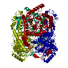
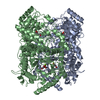
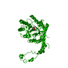
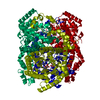
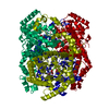
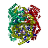
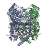
 Streptomyces rubiginosus (bacteria)
 X-RAY DIFFRACTION / NUCLEAR REACTOR / Resolution: 2.19 Å
  Authors
 Citation
 Structure visualization
 Downloads & links
 Other downloads
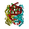
 PDBj
PDBj


 Assembly

 Type: D-saccharide, alpha linking / Mass: 180.156 Da / Num. of mol.: 1
 Mass: 54.938 Da / Num. of mol.: 2 / Source method: obtained synthetically / Formula: Mn
 Mass: 24.305 Da / Num. of mol.: 1 / Source method: obtained synthetically / Formula: Mg
 Mass: 18.015 Da / Num. of mol.: 289 / Source method: isolated from a natural source / Formula: D2O
 Sample preparation
 Processing