Mass: 18.015 Da / Num. of mol.: 320 / Source method: isolated from a natural source / Formula: H2O
Has protein modification
Y
Sequence details
THIS CONSTRUCT WAS EXPRESSED WITH AN N-TERMINAL PURIFICATION TAG MGSDKIHHHHHHENLYFQG. THE TAG WAS ...THIS CONSTRUCT WAS EXPRESSED WITH AN N-TERMINAL PURIFICATION TAG MGSDKIHHHHHHENLYFQG. THE TAG WAS REMOVED WITH TEV PROTEASE LEAVING ONLY A GLYCINE (0) FOLLOWED BY RESIDUES 86-174 OF THE TARGET SEQUENCE - ISOFORM 2 OF FUSE-BINDING PROTEIN 1, UNIPROT ENTRY Q96AE4-2.
-
Experimental details
-
Experiment
Experiment
Method: X-RAY DIFFRACTION / Number of used crystals: 1
-
Sample preparation
Crystal
Density Matthews: 2.66 Å3/Da / Density % sol: 53.84 %
Crystal grow
Temperature: 277 K / Method: vapor diffusion, sitting drop / pH: 8.5 Details: 20.00% Glycerol, 1.60M ammonium dihydrogen phosphate, 0.1M TRIS pH 8.5, NANODROP, VAPOR DIFFUSION, SITTING DROP, temperature 277K
Resolution: 1.8→61.384 Å / Num. obs: 24100 / % possible obs: 78.6 % / Observed criterion σ(I): -3 / Biso Wilson estimate: 14.277 Å2 / Rmerge(I) obs: 0.143 / Net I/σ(I): 9.17
Reflection shell
Resolution (Å)
Rmerge(I) obs
Mean I/σ(I) obs
Num. measured obs
Num. unique obs
Diffraction-ID
% possible all
1.8-1.85
0.923
1.6
15357
4174
1
99.4
1.85-1.9
0.848
1.8
13228
3628
1
88.4
1.9-1.95
0.633
1
289
186
1
4.6
1.95-2.01
0.508
3
14693
3877
1
99.7
2.01-2.08
0.394
3.7
5877
1650
1
44.1
2.08-2.15
0.321
4.5
9110
2497
1
68.8
2.15-2.23
0.324
4.8
12823
3420
1
96.6
2.23-2.32
0.226
6.1
4208
1166
1
34.8
2.32-2.43
0.205
7.1
12487
3230
1
99.8
2.43-2.55
0.194
7.4
11955
3096
1
99.9
2.55-2.68
0.169
8.3
7815
2087
1
70.8
2.68-2.85
0.113
11.6
9171
2427
1
87.6
2.85-3.04
0.093
14
10002
2607
1
100
3.04-3.29
0.079
16.4
9341
2442
1
100
3.29-3.6
0.068
17.8
6603
1781
1
79.5
3.6-4.02
0.059
19.5
4975
1391
1
68.1
4.02-4.65
0.046
23.7
6573
1773
1
99.7
4.65-5.69
0.052
21.4
5780
1502
1
99.7
5.69-8.05
0.051
20.9
4555
1168
1
99.9
8.05-61.384
0.03
29.7
2418
634
1
99.8
-
Phasing
Phasing
Method: MAD
-
Processing
Software
Name
Version
Classification
NB
MolProbity
3beta29
modelbuilding
PDB_EXTRACT
3.1
dataextraction
SOLVE
phasing
XSCALE
July4, 2012
datascaling
REFMAC
5.7.0032
refinement
XDS
datareduction
Refinement
Method to determine structure: MAD / Resolution: 1.8→61.384 Å / Cor.coef. Fo:Fc: 0.95 / Cor.coef. Fo:Fc free: 0.934 / Occupancy max: 1 / Occupancy min: 0.3 / SU B: 4.119 / SU ML: 0.066 / Cross valid method: THROUGHOUT / σ(F): 0 / ESU R: 0.133 / ESU R Free: 0.122 Stereochemistry target values: MAXIMUM LIKELIHOOD WITH PHASES Details: 1. HYDROGENS HAVE BEEN ADDED IN THE RIDING POSITIONS. 2. ATOM RECORDS CONTAIN SUM OF TLS AND RESIDUAL B FACTORS. 3. ANISOU RECORDS CONTAIN SUM OF TLS AND RESIDUAL U FACTORS. 4. WATERS WERE ...Details: 1. HYDROGENS HAVE BEEN ADDED IN THE RIDING POSITIONS. 2. ATOM RECORDS CONTAIN SUM OF TLS AND RESIDUAL B FACTORS. 3. ANISOU RECORDS CONTAIN SUM OF TLS AND RESIDUAL U FACTORS. 4. WATERS WERE EXCLUDED FROM AUTOMATIC TLS ASSIGNMENT. 5. A MET-INHIBITION PROTOCOL WAS USED FOR SELENOMETHIONINE INCORPORATION DURING PROTEIN EXPRESSION. THE OCCUPANCY OF THE SE ATOMS IN THE MSE RESIDUES WAS REDUCED TO 0.75 FOR THE REDUCED SCATTERING POWER DUE TO PARTIAL S-MET INCORPORATION. 6. PHOSPHATE (PO4) MOLECULES FROM THE CRYSTALLIZATION SOLUTION ARE MODELED. 7. DUE TO STRONG ICE RINGS, REFLECTIONS WERE OMITTED IN THE 3.93-3.87, 3.70-3.64, 2.29-2.23, AND 1.95-1.89 RESOLUTION SHELLS LOWERING THE OVERALL COMPLETENESS TO 79.2%. THE NOMINAL RESOLUTION OF THE RESULTING DATASET IS 1.95 A WITH 4124 OBSERVED REFLECTIONS BETWEEN 1.95-1.80 (64.8% COMPLETE FOR THIS SHELL) INCLUDED IN THE REFINEMENT.
Rfactor
Num. reflection
% reflection
Selection details
Rfree
0.1992
1228
5.1 %
RANDOM
Rwork
0.1716
-
-
-
obs
0.173
24098
79.19 %
-
Solvent computation
Ion probe radii: 0.8 Å / Shrinkage radii: 0.8 Å / VDW probe radii: 1.2 Å / Solvent model: BABINET MODEL WITH MASK
In the structure databanks used in Yorodumi, some data are registered as the other names, "COVID-19 virus" and "2019-nCoV". Here are the details of the virus and the list of structure data.
Jan 31, 2019. EMDB accession codes are about to change! (news from PDBe EMDB page)
EMDB accession codes are about to change! (news from PDBe EMDB page)
The allocation of 4 digits for EMDB accession codes will soon come to an end. Whilst these codes will remain in use, new EMDB accession codes will include an additional digit and will expand incrementally as the available range of codes is exhausted. The current 4-digit format prefixed with “EMD-” (i.e. EMD-XXXX) will advance to a 5-digit format (i.e. EMD-XXXXX), and so on. It is currently estimated that the 4-digit codes will be depleted around Spring 2019, at which point the 5-digit format will come into force.
The EM Navigator/Yorodumi systems omit the EMD- prefix.
Related info.:Q: What is EMD? / ID/Accession-code notation in Yorodumi/EM Navigator
Yorodumi is a browser for structure data from EMDB, PDB, SASBDB, etc.
This page is also the successor to EM Navigator detail page, and also detail information page/front-end page for Omokage search.
The word "yorodu" (or yorozu) is an old Japanese word meaning "ten thousand". "mi" (miru) is to see.
Related info.:EMDB / PDB / SASBDB / Comparison of 3 databanks / Yorodumi Search / Aug 31, 2016. New EM Navigator & Yorodumi / Yorodumi Papers / Jmol/JSmol / Function and homology information / Changes in new EM Navigator and Yorodumi
 Movie
Movie Controller

 Yorodumi
 Open data
 Basic information
 Components
 Keywords
 Function and homology information
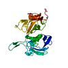
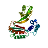
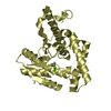
 Homo sapiens (human)
 X-RAY DIFFRACTION /
  Authors
 Citation
 Structure visualization
 Downloads & links
 Other downloads


 PDBj
PDBj
 Assembly
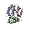


 Mass: 94.971 Da / Num. of mol.: 2 / Source method: obtained synthetically / Formula: PO4
 Mass: 18.015 Da / Num. of mol.: 320 / Source method: isolated from a natural source / Formula: H2O
 Sample preparation
 / Beamline: 8.2.2 / Wavelength: 0.9537,0.9796,0.9793
 Processing