 Movie
Movie Controller

[English] 日本語
 Yorodumi
- PDB-4lby: Identifying ligand binding hot spots in proteins using brominated... -
+ Open data

- Basic information
| Entry | Database: PDB / ID: 4lby | ||||||
|---|---|---|---|---|---|---|---|
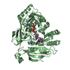
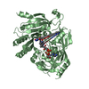
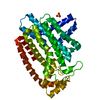
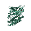
| Title | Identifying ligand binding hot spots in proteins using brominated fragments | ||||||
 Components Components | Elongation factor Tu-A | ||||||
 Keywords Keywords | PROTEIN BINDING / GTPase | ||||||
| Function / homology |  Function and homology information Function and homology informationprotein-synthesizing GTPase / translation elongation factor activity / GTPase activity / GTP binding / magnesium ion binding / cytosol Similarity search - Function | ||||||
| Biological species |   Thermus thermophilus (bacteria) Thermus thermophilus (bacteria) | ||||||
| Method |  X-RAY DIFFRACTION / MOLECULAR REPLACEMENT / molecular replacement / Resolution: 2.692 Å X-RAY DIFFRACTION / MOLECULAR REPLACEMENT / molecular replacement / Resolution: 2.692 Å | ||||||
 Authors Authors | Groftehauge, M.K. / Therkelsen, M. / Taaning, R. / Skrydstrup, T. / Morth, J.P. / Nissen, P. | ||||||
 Citation Citation | Journal: Acta Crystallogr.,Sect.F / Year: 2013 Title: Identifying ligand-binding hot spots in proteins using brominated fragments. Authors: Grftehauge, M.K. / Therkelsen, M.O. / Taaning, R. / Skrydstrup, T. / Morth, J.P. / Nissen, P. | ||||||
| History |
|
- Structure visualization
| Structure viewer | Molecule: MolmilJmol/JSmol |
|---|
- Downloads & links
-Download
| PDBx/mmCIF format | 4lby.cif.gz | 173.4 KB | Display | PDBx/mmCIF format |
|---|---|---|---|---|
| PDB format | pdb4lby.ent.gz | 136.8 KB | Display | PDB format |
| PDBx/mmJSON format | 4lby.json.gz | Tree view | PDBx/mmJSON format | |
| Others |  Other downloads Other downloads |
-Validation report
| Arichive directory | https://data.pdbj.org/pub/pdb/validation_reports/lb/4lbyftp://data.pdbj.org/pub/pdb/validation_reports/lb/4lby | HTTPS FTP |
|---|
-Related structure data
| Related structure data |  4h9gSC  4lbvC  4lbwC  4lbzC  4lc0C C: citing same article ( S: Starting model for refinement |
|---|---|
| Similar structure data |


-Links
 PDBj
PDBj


- Assembly
| Deposited unit | 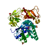
| ||||||||
|---|---|---|---|---|---|---|---|---|---|
| 1 |
| ||||||||
| Unit cell |
|
-Components
-Protein , 1 types, 1 molecules A
| #1: Protein | Mass: 44610.758 Da / Num. of mol.: 1 Source method: isolated from a genetically manipulated source Source: (gene. exp.) Thermus thermophilus (bacteria) / Gene: tuf, tufA / Plasmid: pKK223-3 / Production host: |
|---|
-Non-polymers , 5 types, 87 molecules 


| #2: Chemical | ChemComp-GNP /  Mass: 522.196 Da / Num. of mol.: 1 / Source method: obtained synthetically / Formula: C10H17N6O13P3 Mass: 522.196 Da / Num. of mol.: 1 / Source method: obtained synthetically / Formula: C10H17N6O13P3Comment: GppNHp, GMPPNP, energy-carrying molecule analogue*YM | ||
|---|---|---|---|
| #3: Chemical | ChemComp-MG /  Mass: 24.305 Da / Num. of mol.: 1 / Source method: obtained synthetically / Formula: Mg Mass: 24.305 Da / Num. of mol.: 1 / Source method: obtained synthetically / Formula: Mg | ||
| #4: Chemical | ChemComp-NH4 /  Mass: 18.038 Da / Num. of mol.: 1 / Source method: obtained synthetically / Formula: H4N Mass: 18.038 Da / Num. of mol.: 1 / Source method: obtained synthetically / Formula: H4N | ||
| #5: Chemical | ChemComp-SO4 /  Mass: 96.063 Da / Num. of mol.: 6 / Source method: obtained synthetically / Formula: SO4 Mass: 96.063 Da / Num. of mol.: 6 / Source method: obtained synthetically / Formula: SO4#6: Water | ChemComp-HOH / | Mass: 18.015 Da / Num. of mol.: 78 / Source method: isolated from a natural source / Formula: H2O |
-Experimental details
-Experiment
| Experiment | Method: X-RAY DIFFRACTION / Number of used crystals: 1 |
|---|
- Sample preparation
| Crystal | Density Matthews: 3.21 Å3/Da / Density % sol: 61.65 % |
|---|---|
| Crystal grow | Temperature: 292 K / Method: vapor diffusion, sitting drop / pH: 7.6 Details: 1.8M ammonium sulfate, 15% sucrose, 0.1M Tris-HCl, pH 7.6, VAPOR DIFFUSION, SITTING DROP, temperature 292K |
-Data collection
| Diffraction | Mean temperature: 100 K |
|---|---|
| Diffraction source | Source: ROTATING ANODE / Type: ENRAF-NONIUS FR571 / Wavelength: 1.5418 Å |
| Detector | Type: MAR scanner 345 mm plate / Detector: IMAGE PLATE |
| Radiation | Protocol: SINGLE WAVELENGTH / Monochromatic (M) / Laue (L): M / Scattering type: x-ray |
| Radiation wavelength | Wavelength: 1.5418 Å / Relative weight: 1 |
| Reflection | Resolution: 2.69→33.35 Å / Num. obs: 15665 / % possible obs: 95.1 % / Redundancy: 1.8 % / Rmerge(I) obs: 0.115 / Net I/σ(I): 12.6 |
| Reflection shell | Resolution: 2.69→2.76 Å / Redundancy: 1.8 % / Rmerge(I) obs: 0.639 / Mean I/σ(I) obs: 2.3 / Num. measured all: 4290 / Num. unique all: 1150 / % possible all: 94.8 |
-Phasing
| Phasing | Method: molecular replacement |
|---|
- Processing
| Software |
| ||||||||||||||||||||||||||||||||||||||||||||||||||||||||||||||||||||||||||||||||||||||||||||||||||||
|---|---|---|---|---|---|---|---|---|---|---|---|---|---|---|---|---|---|---|---|---|---|---|---|---|---|---|---|---|---|---|---|---|---|---|---|---|---|---|---|---|---|---|---|---|---|---|---|---|---|---|---|---|---|---|---|---|---|---|---|---|---|---|---|---|---|---|---|---|---|---|---|---|---|---|---|---|---|---|---|---|---|---|---|---|---|---|---|---|---|---|---|---|---|---|---|---|---|---|---|---|---|
| Refinement | Method to determine structure: MOLECULAR REPLACEMENT Starting model: pdb entry 4H9G Resolution: 2.692→33.35 Å / Occupancy max: 1 / Occupancy min: 0.73 / SU ML: 0.3 / σ(F): 1.37 / Phase error: 19.91 / Stereochemistry target values: ML
| ||||||||||||||||||||||||||||||||||||||||||||||||||||||||||||||||||||||||||||||||||||||||||||||||||||
| Solvent computation | Shrinkage radii: 0.9 Å / VDW probe radii: 1.11 Å / Solvent model: FLAT BULK SOLVENT MODEL | ||||||||||||||||||||||||||||||||||||||||||||||||||||||||||||||||||||||||||||||||||||||||||||||||||||
| Displacement parameters | Biso max: 115.65 Å2 / Biso mean: 41.5332 Å2 / Biso min: 7.39 Å2 | ||||||||||||||||||||||||||||||||||||||||||||||||||||||||||||||||||||||||||||||||||||||||||||||||||||
| Refinement step | Cycle: LAST / Resolution: 2.692→33.35 Å
| ||||||||||||||||||||||||||||||||||||||||||||||||||||||||||||||||||||||||||||||||||||||||||||||||||||
| Refine LS restraints |
| ||||||||||||||||||||||||||||||||||||||||||||||||||||||||||||||||||||||||||||||||||||||||||||||||||||
| LS refinement shell | Refine-ID: X-RAY DIFFRACTION / Total num. of bins used: 6 / % reflection obs: 100 %
| ||||||||||||||||||||||||||||||||||||||||||||||||||||||||||||||||||||||||||||||||||||||||||||||||||||
| Refinement TLS params. | Method: refined / Refine-ID: X-RAY DIFFRACTION
| ||||||||||||||||||||||||||||||||||||||||||||||||||||||||||||||||||||||||||||||||||||||||||||||||||||
| Refinement TLS group |
|
