 Movie
Movie Controller

[English] 日本語
 Yorodumi
- PDB-5qqc: Crystal Structure of T. cruzi FPPS after initial refinement with ... -
+ Open data

- Basic information
| Entry | Database: PDB / ID: 5qqc | ||||||
|---|---|---|---|---|---|---|---|
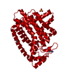
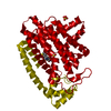
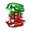
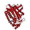
| Title | Crystal Structure of T. cruzi FPPS after initial refinement with no ligand modelled (structure $n) | ||||||
 Components Components | Farnesyl diphosphate synthase | ||||||
 Keywords Keywords | TRANSFERASE / SGC - Diamond I04-1 fragment screening / PanDDA / XChemExplorer | ||||||
| Function / homology |  Function and homology information Function and homology informationtrans, trans-farnesyl diphosphate biosynthetic process / dimethylallyltranstransferase activity / (2E,6E)-farnesyl diphosphate synthase activity / metal ion binding / cytoplasm Similarity search - Function | ||||||
| Biological species |  | ||||||
| Method |  X-RAY DIFFRACTION / SYNCHROTRON / FOURIER SYNTHESIS / molecular replacement / Resolution: 1.62 Å X-RAY DIFFRACTION / SYNCHROTRON / FOURIER SYNTHESIS / molecular replacement / Resolution: 1.62 Å | ||||||
 Authors Authors | Petrick, J.K. / Nelson, E.R. / Muenzker, L. / Krojer, T. / Douangamath, A. / Brandao-Neto, J. / von Delft, F. / Dekker, C. / Jahnke, W. | ||||||
 Citation Citation | Journal: To Be Published Title: PanDDA analysis group deposition of ground-state model - FPPS screened against the DSI Fragment Library Authors: Petrick, J.K. / Muenzker, L. / von Delft, F. / Jahnke, W. | ||||||
| History |
|
- Structure visualization
| Structure viewer | Molecule: MolmilJmol/JSmol |
|---|
- Downloads & links
-Download
| PDBx/mmCIF format | 5qqc.cif.gz | 92.5 KB | Display | PDBx/mmCIF format |
|---|---|---|---|---|
| PDB format | pdb5qqc.ent.gz | 68.6 KB | Display | PDB format |
| PDBx/mmJSON format | 5qqc.json.gz | Tree view | PDBx/mmJSON format | |
| Others |  Other downloads Other downloads |
-Validation report
| Arichive directory | https://data.pdbj.org/pub/pdb/validation_reports/qq/5qqcftp://data.pdbj.org/pub/pdb/validation_reports/qq/5qqc | HTTPS FTP |
|---|
-Group deposition
| ID | G_1002064 (1 entries) |
|---|---|
| Title | PanDDA analysis group deposition of ground-state model |
| Type | ground state |
| Description | FPPS screened against the DSI Fragment Library by X-ray Crystallography at the XChem facility of Diamond Light Source beamline I04-1 |
-Related structure data
| Related structure data |  1yhkS S: Starting model for refinement |
|---|---|
| Similar structure data |


-Links
 PDBj
PDBj
- Assembly
| Deposited unit | 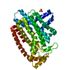
| ||||||||
|---|---|---|---|---|---|---|---|---|---|
| 1 | 
| ||||||||
| Unit cell |
|
-Components
| #1: Protein | Mass: 41359.574 Da / Num. of mol.: 1 Source method: isolated from a genetically manipulated source Source: (gene. exp.)  | ||||||||
|---|---|---|---|---|---|---|---|---|---|
| #2: Chemical |   Mass: 96.063 Da / Num. of mol.: 2 / Source method: obtained synthetically / Formula: SO4 Mass: 96.063 Da / Num. of mol.: 2 / Source method: obtained synthetically / Formula: SO4#3: Chemical | ChemComp-ACT / |   Mass: 59.044 Da / Num. of mol.: 1 / Source method: obtained synthetically / Formula: C2H3O2 Mass: 59.044 Da / Num. of mol.: 1 / Source method: obtained synthetically / Formula: C2H3O2#4: Chemical |   Mass: 24.305 Da / Num. of mol.: 2 / Source method: obtained synthetically / Formula: Mg Mass: 24.305 Da / Num. of mol.: 2 / Source method: obtained synthetically / Formula: Mg#5: Water | ChemComp-HOH / |  Mass: 18.015 Da / Num. of mol.: 291 / Source method: isolated from a natural source / Formula: H2O Mass: 18.015 Da / Num. of mol.: 291 / Source method: isolated from a natural source / Formula: H2OHas protein modification | N | |
-Experimental details
-Experiment
| Experiment | Method: X-RAY DIFFRACTION / Number of used crystals: 1 |
|---|
- Sample preparation
| Crystal | Density Matthews: 2.3 Å3/Da / Density % sol: 46.57 % / Mosaicity: 0 ° |
|---|---|
| Crystal grow | Temperature: 293 K / Method: vapor diffusion, hanging drop / pH: 6.5 Details: 80 mM MES, 4 mM ZnSO4, 12.36% w/v PEG MME 550, 11.57% v/v glycerol |
-Data collection
| Diffraction | Mean temperature: 100 K | |||||||||||||||||||||||||||
|---|---|---|---|---|---|---|---|---|---|---|---|---|---|---|---|---|---|---|---|---|---|---|---|---|---|---|---|---|
| Diffraction source | Source: SYNCHROTRON / Site: Diamond  / Beamline: I04-1 / Wavelength: 0.91587 Å / Beamline: I04-1 / Wavelength: 0.91587 Å | |||||||||||||||||||||||||||
| Detector | Type: DECTRIS PILATUS 6M-F / Detector: PIXEL / Date: Oct 8, 2017 | |||||||||||||||||||||||||||
| Radiation | Protocol: SINGLE WAVELENGTH / Scattering type: x-ray | |||||||||||||||||||||||||||
| Radiation wavelength | Wavelength: 0.91587 Å / Relative weight: 1 | |||||||||||||||||||||||||||
| Reflection | Resolution: 1.62→132.19 Å / Num. obs: 51799 / % possible obs: 100 % / Redundancy: 17.7 % / CC1/2: 0.999 / Rmerge(I) obs: 0.1 / Rpim(I) all: 0.024 / Rrim(I) all: 0.103 / Net I/σ(I): 15 / Num. measured all: 915855 / Scaling rejects: 434 | |||||||||||||||||||||||||||
| Reflection shell | Diffraction-ID: 1 / % possible all: 100
|
-Phasing
| Phasing | Method: molecular replacement |
|---|
- Processing
| Software |
| |||||||||||||||||||||||||||||||||||||||||||||||||||||||||||||||||||||||||||
|---|---|---|---|---|---|---|---|---|---|---|---|---|---|---|---|---|---|---|---|---|---|---|---|---|---|---|---|---|---|---|---|---|---|---|---|---|---|---|---|---|---|---|---|---|---|---|---|---|---|---|---|---|---|---|---|---|---|---|---|---|---|---|---|---|---|---|---|---|---|---|---|---|---|---|---|---|
| Refinement | Method to determine structure: FOURIER SYNTHESIS Starting model: 1YHK Resolution: 1.62→66.09 Å / Cor.coef. Fo:Fc: 0.968 / Cor.coef. Fo:Fc free: 0.955 / SU B: 2.377 / SU ML: 0.077 / Cross valid method: THROUGHOUT / σ(F): 0 / ESU R: 0.094 / ESU R Free: 0.096 / Stereochemistry target values: MAXIMUM LIKELIHOOD Details: HYDROGENS HAVE BEEN ADDED IN THE RIDING POSITIONS U VALUES : REFINED INDIVIDUALLY
| |||||||||||||||||||||||||||||||||||||||||||||||||||||||||||||||||||||||||||
| Solvent computation | Ion probe radii: 0.8 Å / Shrinkage radii: 0.8 Å / VDW probe radii: 1.2 Å / Solvent model: MASK | |||||||||||||||||||||||||||||||||||||||||||||||||||||||||||||||||||||||||||
| Displacement parameters | Biso max: 102.74 Å2 / Biso mean: 30.73 Å2 / Biso min: 14.89 Å2
| |||||||||||||||||||||||||||||||||||||||||||||||||||||||||||||||||||||||||||
| Refinement step | Cycle: final / Resolution: 1.62→66.09 Å
| |||||||||||||||||||||||||||||||||||||||||||||||||||||||||||||||||||||||||||
| Refine LS restraints |
| |||||||||||||||||||||||||||||||||||||||||||||||||||||||||||||||||||||||||||
| LS refinement shell | Resolution: 1.62→1.662 Å / Total num. of bins used: 20
|
