 Movie
Movie Controller

[English] 日本語
 Yorodumi
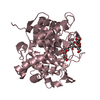
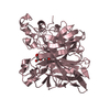
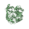
- PDB-4lbh: 5-chloro-2-hydroxyhydroquinone dehydrochlorinase (TftG) from Burk... -
+ Open data

- Basic information
| Entry | Database: PDB / ID: 4lbh | ||||||
|---|---|---|---|---|---|---|---|
| Title | 5-chloro-2-hydroxyhydroquinone dehydrochlorinase (TftG) from Burkholderia phenoliruptrix AC1100: Apo-form | ||||||
 Components Components | 5-chloro-2-hydroxyhydroquinone dehydrochlorinase (TftG) | ||||||
 Keywords Keywords | LYASE | ||||||
| Function / homology |  Function and homology information Function and homology information: / YCII-related / YCII-related domain / Dimeric alpha+beta barrel / Dimeric alpha-beta barrel / Alpha-Beta Plaits / 2-Layer Sandwich / Alpha Beta Similarity search - Domain/homology | ||||||
| Biological species |  Burkholderia cepacia (bacteria) Burkholderia cepacia (bacteria) | ||||||
| Method |  X-RAY DIFFRACTION / SYNCHROTRON / MOLECULAR REPLACEMENT / Resolution: 1.75 Å X-RAY DIFFRACTION / SYNCHROTRON / MOLECULAR REPLACEMENT / Resolution: 1.75 Å | ||||||
 Authors Authors | Hayes, R.P. / Lewis, K.M. / Xun, L. / Kang, C. | ||||||
 Citation Citation | Journal: J.Biol.Chem. / Year: 2013 Title: Catalytic Mechanism of 5-Chlorohydroxyhydroquinone Dehydrochlorinase from the YCII Superfamily of Largely Unknown Function. Authors: Hayes, R.P. / Lewis, K.M. / Xun, L. / Kang, C. | ||||||
| History |
|
- Structure visualization
| Structure viewer | Molecule: MolmilJmol/JSmol |
|---|
- Downloads & links
-Download
| PDBx/mmCIF format | 4lbh.cif.gz | 33.1 KB | Display | PDBx/mmCIF format |
|---|---|---|---|---|
| PDB format | pdb4lbh.ent.gz | 22 KB | Display | PDB format |
| PDBx/mmJSON format | 4lbh.json.gz | Tree view | PDBx/mmJSON format | |
| Others |  Other downloads Other downloads |
-Validation report
| Arichive directory | https://data.pdbj.org/pub/pdb/validation_reports/lb/4lbhftp://data.pdbj.org/pub/pdb/validation_reports/lb/4lbh | HTTPS FTP |
|---|
-Related structure data


-Links
 PDBj
PDBj- Assembly
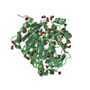
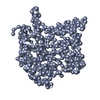
| Deposited unit | 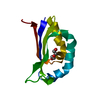
| ||||||||||||
|---|---|---|---|---|---|---|---|---|---|---|---|---|---|
| 1 | 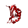
| ||||||||||||
| Unit cell |
| ||||||||||||
| Components on special symmetry positions |
|
-Components
| #1: Protein | Mass: 11181.664 Da / Num. of mol.: 1 Source method: isolated from a genetically manipulated source Source: (gene. exp.) Burkholderia cepacia (bacteria) / Production host: |
|---|---|
| #2: Chemical | ChemComp-PEG /   Mass: 106.120 Da / Num. of mol.: 1 / Source method: obtained synthetically / Formula: C4H10O3 Mass: 106.120 Da / Num. of mol.: 1 / Source method: obtained synthetically / Formula: C4H10O3 |
| #3: Water | ChemComp-HOH /  Mass: 18.015 Da / Num. of mol.: 79 / Source method: isolated from a natural source / Formula: H2O Mass: 18.015 Da / Num. of mol.: 79 / Source method: isolated from a natural source / Formula: H2O |
-Experimental details
-Experiment
| Experiment | Method: X-RAY DIFFRACTION / Number of used crystals: 1 |
|---|
- Sample preparation
| Crystal | Density Matthews: 2.7 Å3/Da / Density % sol: 54.42 % |
|---|---|
| Crystal grow | Temperature: 277.15 K / Method: vapor diffusion, hanging drop / pH: 7.5 Details: 30% (w/v) polyethylene glycol 1500, pH 7.5, VAPOR DIFFUSION, HANGING DROP, temperature 277.15K |
-Data collection
| Diffraction | Mean temperature: 100 K | ||||||||||||||||||
|---|---|---|---|---|---|---|---|---|---|---|---|---|---|---|---|---|---|---|---|
| Diffraction source | Source: SYNCHROTRON / Site: ALS  / Beamline: 8.2.1 / Wavelength: 1 Å / Beamline: 8.2.1 / Wavelength: 1 Å | ||||||||||||||||||
| Detector | Type: ADSC QUANTUM 315r / Detector: CCD / Date: Jan 28, 2013 | ||||||||||||||||||
| Radiation | Monochromator: Double crystal, Si(111) / Protocol: SINGLE WAVELENGTH / Monochromatic (M) / Laue (L): M / Scattering type: x-ray | ||||||||||||||||||
| Radiation wavelength | Wavelength: 1 Å / Relative weight: 1 | ||||||||||||||||||
| Reflection | Resolution: 1.75→50 Å / Num. all: 12883 / Num. obs: 12883 / % possible obs: 99.6 % / Observed criterion σ(I): -3 | ||||||||||||||||||
| Reflection shell |
|
- Processing
| Software |
| ||||||||||||||||||||||||||||||||||||||||||||||||||||||||||||||||||||||
|---|---|---|---|---|---|---|---|---|---|---|---|---|---|---|---|---|---|---|---|---|---|---|---|---|---|---|---|---|---|---|---|---|---|---|---|---|---|---|---|---|---|---|---|---|---|---|---|---|---|---|---|---|---|---|---|---|---|---|---|---|---|---|---|---|---|---|---|---|---|---|---|
| Refinement | Method to determine structure: MOLECULAR REPLACEMENT / Resolution: 1.75→45.158 Å / SU ML: 0.15 / σ(F): 1.34 / Phase error: 23.43 / Stereochemistry target values: ML
| ||||||||||||||||||||||||||||||||||||||||||||||||||||||||||||||||||||||
| Solvent computation | Shrinkage radii: 0.9 Å / VDW probe radii: 1.11 Å / Solvent model: FLAT BULK SOLVENT MODEL | ||||||||||||||||||||||||||||||||||||||||||||||||||||||||||||||||||||||
| Refinement step | Cycle: LAST / Resolution: 1.75→45.158 Å
| ||||||||||||||||||||||||||||||||||||||||||||||||||||||||||||||||||||||
| Refine LS restraints |
| ||||||||||||||||||||||||||||||||||||||||||||||||||||||||||||||||||||||
| LS refinement shell |
|
