 Movie
Movie Controller

[English] 日本語
 Yorodumi
- PDB-4l98: Crystal structure of the complex of F360L PPARgamma mutant with t... -
+ Open data

- Basic information
| Entry | Database: PDB / ID: 4l98 | ||||||
|---|---|---|---|---|---|---|---|
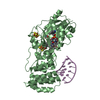
| Title | Crystal structure of the complex of F360L PPARgamma mutant with the ligand LT175 | ||||||
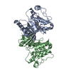
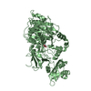
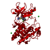
 Components Components | Peroxisome proliferator-activated receptor gamma | ||||||
 Keywords Keywords | TRANSCRIPTION/TRANSCRIPTION inhibitor / transcription factor / RXRalpha / TRANSCRIPTION-TRANSCRIPTION inhibitor complex | ||||||
| Function / homology |  Function and homology information Function and homology informationprostaglandin receptor activity / negative regulation of receptor signaling pathway via STAT / MECP2 regulates transcription factors / beige fat cell differentiation / negative regulation of vascular endothelial cell proliferation / negative regulation of extracellular matrix assembly / negative regulation of connective tissue replacement involved in inflammatory response wound healing / positive regulation of cholesterol transport / negative regulation of cellular response to transforming growth factor beta stimulus / arachidonate binding ...prostaglandin receptor activity / negative regulation of receptor signaling pathway via STAT / MECP2 regulates transcription factors / beige fat cell differentiation / negative regulation of vascular endothelial cell proliferation / negative regulation of extracellular matrix assembly / negative regulation of connective tissue replacement involved in inflammatory response wound healing / positive regulation of cholesterol transport / negative regulation of cellular response to transforming growth factor beta stimulus / arachidonate binding / positive regulation of adiponectin secretion / DNA binding domain binding / negative regulation of cardiac muscle hypertrophy in response to stress / positive regulation of vascular associated smooth muscle cell apoptotic process / positive regulation of lipid metabolic process / positive regulation of fatty acid metabolic process / STAT family protein binding / WW domain binding / negative regulation of type II interferon-mediated signaling pathway / LBD domain binding / negative regulation of cholesterol storage / response to lipid / positive regulation of lipoprotein transport / negative regulation of SMAD protein signal transduction / lipid homeostasis / E-box binding / R-SMAD binding / negative regulation of blood vessel endothelial cell migration / white fat cell differentiation / alpha-actinin binding / negative regulation of vascular associated smooth muscle cell proliferation / negative regulation of macrophage derived foam cell differentiation / negative regulation of lipid storage / positive regulation of cholesterol efflux / negative regulation of BMP signaling pathway / monocyte differentiation / cell fate commitment / cellular response to low-density lipoprotein particle stimulus / long-chain fatty acid transport / BMP signaling pathway / negative regulation of mitochondrial fission / negative regulation of osteoblast differentiation / positive regulation of fat cell differentiation / nuclear retinoid X receptor binding / fat cell differentiation / Transcriptional regulation of brown and beige adipocyte differentiation by EBF2 / retinoic acid receptor signaling pathway / intracellular receptor signaling pathway / negative regulation of MAPK cascade / peptide binding / peroxisome proliferator activated receptor signaling pathway / cell maturation / epithelial cell differentiation / hormone-mediated signaling pathway / regulation of cellular response to insulin stimulus / positive regulation of adipose tissue development / response to nutrient / negative regulation of miRNA transcription / brown fat cell differentiation / negative regulation of angiogenesis / placenta development / Regulation of PTEN gene transcription / transcription coregulator binding / SUMOylation of intracellular receptors / positive regulation of apoptotic signaling pathway / negative regulation of smooth muscle cell proliferation / negative regulation of transforming growth factor beta receptor signaling pathway / PPARA activates gene expression / fatty acid metabolic process / Nuclear Receptor transcription pathway / Transcriptional regulation of white adipocyte differentiation / regulation of circadian rhythm / positive regulation of miRNA transcription / mRNA transcription by RNA polymerase II / DNA-binding transcription repressor activity, RNA polymerase II-specific / nuclear receptor activity / negative regulation of inflammatory response / regulation of blood pressure / RNA polymerase II transcription regulator complex / cellular response to insulin stimulus / rhythmic process / glucose homeostasis / MLL4 and MLL3 complexes regulate expression of PPARG target genes in adipogenesis and hepatic steatosis / double-stranded DNA binding / DNA-binding transcription activator activity, RNA polymerase II-specific / cellular response to hypoxia / DNA-binding transcription factor binding / sequence-specific DNA binding / nucleic acid binding / DNA-binding transcription factor activity, RNA polymerase II-specific / cell differentiation / signaling receptor complex / transcription cis-regulatory region binding / RNA polymerase II cis-regulatory region sequence-specific DNA binding / DNA-binding transcription factor activity / negative regulation of gene expression / innate immune response / negative regulation of DNA-templated transcription / chromatin binding / positive regulation of gene expression Similarity search - Function | ||||||
| Biological species |  Homo sapiens (human) Homo sapiens (human) | ||||||
| Method |  X-RAY DIFFRACTION / SYNCHROTRON / MOLECULAR REPLACEMENT / Resolution: 2.28 Å X-RAY DIFFRACTION / SYNCHROTRON / MOLECULAR REPLACEMENT / Resolution: 2.28 Å | ||||||
 Authors Authors | Pochetti, G. / Montanari, R. / Consalvi, V. / Chiaraluce, R. / Pasquo, A. / Capelli, D. / Loiodice, F. / Laghezza, A. / Lori, C. | ||||||
 Citation Citation | Journal: Acta Crystallogr.,Sect.D / Year: 2014 Title: Structural basis of the transactivation deficiency of the human PPAR gamma F360L mutant associated with familial partial lipodystrophy. Authors: Lori, C. / Pasquo, A. / Montanari, R. / Capelli, D. / Consalvi, V. / Chiaraluce, R. / Cervoni, L. / Loiodice, F. / Laghezza, A. / Aschi, M. / Giorgi, A. / Pochetti, G. #1: Journal: J.Med.Chem. / Year: 2008Title: Crystal structure of the peroxisome proliferator-activated receptor gamma (PPARgamma) ligand binding domain complexed with a novel partial agonist: a new region of the hydrophobic pocket could ...Title: Crystal structure of the peroxisome proliferator-activated receptor gamma (PPARgamma) ligand binding domain complexed with a novel partial agonist: a new region of the hydrophobic pocket could be exploited for drug design. Authors: Montanari, R. / Saccoccia, F. / Scotti, E. / Crestani, M. / Godio, C. / Gilardi, F. / Loiodice, F. / Fracchiolla, G. / Laghezza, A. / Tortorella, P. / Lavecchia, A. / Novellino, E. / Mazza, ...Authors: Montanari, R. / Saccoccia, F. / Scotti, E. / Crestani, M. / Godio, C. / Gilardi, F. / Loiodice, F. / Fracchiolla, G. / Laghezza, A. / Tortorella, P. / Lavecchia, A. / Novellino, E. / Mazza, F. / Aschi, M. / Pochetti, G. | ||||||
| History |
|
- Structure visualization
| Structure viewer | Molecule: MolmilJmol/JSmol |
|---|
- Downloads & links
-Download
| PDBx/mmCIF format | 4l98.cif.gz | 219.6 KB | Display | PDBx/mmCIF format |
|---|---|---|---|---|
| PDB format | pdb4l98.ent.gz | 176.3 KB | Display | PDB format |
| PDBx/mmJSON format | 4l98.json.gz | Tree view | PDBx/mmJSON format | |
| Others |  Other downloads Other downloads |
-Validation report
| Arichive directory | https://data.pdbj.org/pub/pdb/validation_reports/l9/4l98ftp://data.pdbj.org/pub/pdb/validation_reports/l9/4l98 | HTTPS FTP |
|---|
-Related structure data
| Related structure data |  4l96C  4o8fC  3b3kS S: Starting model for refinement C: citing same article ( |
|---|---|
| Similar structure data |


-Links
 PDBj
PDBj- Assembly
| Deposited unit | 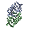
| ||||||||||||||||||
|---|---|---|---|---|---|---|---|---|---|---|---|---|---|---|---|---|---|---|---|
| 1 |
| ||||||||||||||||||
| Unit cell |
| ||||||||||||||||||
| Noncrystallographic symmetry (NCS) | NCS domain:
NCS domain segments: Component-ID: _ / Ens-ID: 1 / Beg auth comp-ID: SER / Beg label comp-ID: SER / End auth comp-ID: LEU / End label comp-ID: LEU / Refine code: _ / Auth seq-ID: 208 - 476 / Label seq-ID: 6 - 274
|
-Components
| #1: Protein | Mass: 31376.473 Da / Num. of mol.: 2 / Fragment: LIGAND BINDING DOMAIN (UNP RESIDUES 235-505) / Mutation: F360L Source method: isolated from a genetically manipulated source Source: (gene. exp.) Homo sapiens (human) / Gene: PPARG, NR1C3 / Plasmid: pET28a / Production host:  #2: Chemical |   Mass: 318.366 Da / Num. of mol.: 2 / Source method: obtained synthetically / Formula: C21H18O3 Mass: 318.366 Da / Num. of mol.: 2 / Source method: obtained synthetically / Formula: C21H18O3#3: Water | ChemComp-HOH / |  Mass: 18.015 Da / Num. of mol.: 227 / Source method: isolated from a natural source / Formula: H2O Mass: 18.015 Da / Num. of mol.: 227 / Source method: isolated from a natural source / Formula: H2O |
|---|
-Experimental details
-Experiment
| Experiment | Method: X-RAY DIFFRACTION / Number of used crystals: 1 |
|---|
- Sample preparation
| Crystal | Density Matthews: 3.4 Å3/Da / Density % sol: 63.84 % |
|---|---|
| Crystal grow | Temperature: 293 K / Method: vapor diffusion, sitting drop / pH: 7 Details: 3.3 M Sodium Formate, pH 7.0, VAPOR DIFFUSION, SITTING DROP, temperature 293K |
-Data collection
| Diffraction | Mean temperature: 100 K |
|---|---|
| Diffraction source | Source: SYNCHROTRON / Site: ESRF  / Beamline: ID23-1 / Wavelength: 0.973 Å / Beamline: ID23-1 / Wavelength: 0.973 Å |
| Detector | Type: PSI PILATUS 6M / Detector: PIXEL / Date: May 1, 2013 |
| Radiation | Monochromator: Si 111 / Protocol: SINGLE WAVELENGTH / Monochromatic (M) / Laue (L): M / Scattering type: x-ray |
| Radiation wavelength | Wavelength: 0.973 Å / Relative weight: 1 |
| Reflection | Resolution: 2.28→50 Å / Num. all: 39713 / Num. obs: 39713 / % possible obs: 99.9 % / Observed criterion σ(F): 0 / Observed criterion σ(I): 0 / Rmerge(I) obs: 0.088 / Net I/σ(I): 18.9 |
| Reflection shell | Resolution: 2.28→2.5 Å / Rmerge(I) obs: 0.424 / Mean I/σ(I) obs: 4.8 / % possible all: 99.9 |
- Processing
| Software |
| ||||||||||||||||||||||||||||||||||||||||||||||||||||||||||||||||||||||||||||||||||||||||||||||||||||||||||||||||||||||||||||||||||||||||||||||||||||||||||||||||||||||||||||||||||||||
|---|---|---|---|---|---|---|---|---|---|---|---|---|---|---|---|---|---|---|---|---|---|---|---|---|---|---|---|---|---|---|---|---|---|---|---|---|---|---|---|---|---|---|---|---|---|---|---|---|---|---|---|---|---|---|---|---|---|---|---|---|---|---|---|---|---|---|---|---|---|---|---|---|---|---|---|---|---|---|---|---|---|---|---|---|---|---|---|---|---|---|---|---|---|---|---|---|---|---|---|---|---|---|---|---|---|---|---|---|---|---|---|---|---|---|---|---|---|---|---|---|---|---|---|---|---|---|---|---|---|---|---|---|---|---|---|---|---|---|---|---|---|---|---|---|---|---|---|---|---|---|---|---|---|---|---|---|---|---|---|---|---|---|---|---|---|---|---|---|---|---|---|---|---|---|---|---|---|---|---|---|---|---|---|
| Refinement | Method to determine structure: MOLECULAR REPLACEMENT Starting model: 3B3K Resolution: 2.28→117.74 Å / Cor.coef. Fo:Fc: 0.933 / Cor.coef. Fo:Fc free: 0.914 / SU B: 10.851 / SU ML: 0.145 / Cross valid method: THROUGHOUT / ESU R: 0.242 / ESU R Free: 0.21 / Stereochemistry target values: MAXIMUM LIKELIHOOD / Details: HYDROGENS HAVE BEEN ADDED IN THE RIDING POSITIONS
| ||||||||||||||||||||||||||||||||||||||||||||||||||||||||||||||||||||||||||||||||||||||||||||||||||||||||||||||||||||||||||||||||||||||||||||||||||||||||||||||||||||||||||||||||||||||
| Solvent computation | Ion probe radii: 1 Å / Shrinkage radii: 1 Å / VDW probe radii: 1.2 Å / Solvent model: MASK | ||||||||||||||||||||||||||||||||||||||||||||||||||||||||||||||||||||||||||||||||||||||||||||||||||||||||||||||||||||||||||||||||||||||||||||||||||||||||||||||||||||||||||||||||||||||
| Displacement parameters | Biso mean: 43.074 Å2
| ||||||||||||||||||||||||||||||||||||||||||||||||||||||||||||||||||||||||||||||||||||||||||||||||||||||||||||||||||||||||||||||||||||||||||||||||||||||||||||||||||||||||||||||||||||||
| Refinement step | Cycle: LAST / Resolution: 2.28→117.74 Å
| ||||||||||||||||||||||||||||||||||||||||||||||||||||||||||||||||||||||||||||||||||||||||||||||||||||||||||||||||||||||||||||||||||||||||||||||||||||||||||||||||||||||||||||||||||||||
| Refine LS restraints |
|