 Movie
Movie Controller

[English] 日本語
 Yorodumi
- PDB-4l95: Crystal structure of gamma glutamyl hydrolase (H218N) from zebrafish -
+ Open data

- Basic information
| Entry | Database: PDB / ID: 4l95 | ||||||
|---|---|---|---|---|---|---|---|
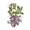
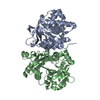
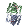
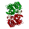
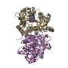
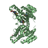
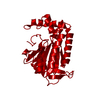
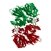
| Title | Crystal structure of gamma glutamyl hydrolase (H218N) from zebrafish | ||||||
 Components Components | Gamma-glutamyl hydrolase | ||||||
 Keywords Keywords | HYDROLASE / Sandwiched-like domain | ||||||
| Function / homology |  Function and homology information Function and homology informationfolate gamma-glutamyl hydrolase / tetrahydrofolylpolyglutamate metabolic process / Neutrophil degranulation / gamma-glutamyl-peptidase activity / folic acid-containing compound metabolic process / vacuole / extracellular region Similarity search - Function | ||||||
| Biological species |  | ||||||
| Method |  X-RAY DIFFRACTION / SYNCHROTRON / MOLECULAR REPLACEMENT / Resolution: 2.34 Å X-RAY DIFFRACTION / SYNCHROTRON / MOLECULAR REPLACEMENT / Resolution: 2.34 Å | ||||||
 Authors Authors | Chuankhayan, P. / Kao, T.-T. / Chen, C.-J. / Fu, T.-F. | ||||||
 Citation Citation | Journal: J.Med.Chem. / Year: 2013 Title: Structural insights into the hydrolysis and polymorphism of methotrexate polyglutamate by zebrafish gamma-glutamyl hydrolase Authors: Chuankhayan, P. / Kao, T.-T. / Lin, C.-C. / Guan, H.-H. / Nakagawa, A. / Fu, T.-F. / Chen, C.-J. | ||||||
| History |
|
- Structure visualization
| Structure viewer | Molecule: MolmilJmol/JSmol |
|---|
- Downloads & links
-Download
| PDBx/mmCIF format | 4l95.cif.gz | 345.6 KB | Display | PDBx/mmCIF format |
|---|---|---|---|---|
| PDB format | pdb4l95.ent.gz | 283 KB | Display | PDB format |
| PDBx/mmJSON format | 4l95.json.gz | Tree view | PDBx/mmJSON format | |
| Others |  Other downloads Other downloads |
-Validation report
| Arichive directory | https://data.pdbj.org/pub/pdb/validation_reports/l9/4l95ftp://data.pdbj.org/pub/pdb/validation_reports/l9/4l95 | HTTPS FTP |
|---|
-Related structure data
| Related structure data |  4l7qC  4l8fC  4l8wC  4l8yC 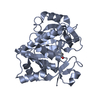 1l9xS C: citing same article ( S: Starting model for refinement |
|---|---|
| Similar structure data |


-Links
 PDBj
PDBj

- Assembly
| Deposited unit | 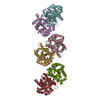
| ||||||||
|---|---|---|---|---|---|---|---|---|---|
| 1 | 
| ||||||||
| 2 | 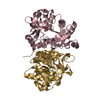
| ||||||||
| 3 | 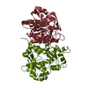
| ||||||||
| Unit cell |
|
-Components
| #1: Protein | Mass: 35392.918 Da / Num. of mol.: 6 / Mutation: H218N Source method: isolated from a genetically manipulated source Source: (gene. exp.)  References: UniProt: Q6NY42, folate gamma-glutamyl hydrolase #2: Chemical |   Mass: 92.094 Da / Num. of mol.: 3 / Source method: obtained synthetically / Formula: C3H8O3 Mass: 92.094 Da / Num. of mol.: 3 / Source method: obtained synthetically / Formula: C3H8O3#3: Water | ChemComp-HOH / |  Mass: 18.015 Da / Num. of mol.: 208 / Source method: isolated from a natural source / Formula: H2O Mass: 18.015 Da / Num. of mol.: 208 / Source method: isolated from a natural source / Formula: H2O |
|---|
-Experimental details
-Experiment
| Experiment | Method: X-RAY DIFFRACTION / Number of used crystals: 1 |
|---|
- Sample preparation
| Crystal | Density Matthews: 2.52 Å3/Da / Density % sol: 51.13 % |
|---|---|
| Crystal grow | Temperature: 291 K / Method: vapor diffusion, hanging drop / pH: 6.2 Details: 10%(w/v) PEG 8000, 0.2M NaCl, 0.1M Na/K buffer, pH 6.2, VAPOR DIFFUSION, HANGING DROP, temperature 291K |
-Data collection
| Diffraction | Mean temperature: 110 K |
|---|---|
| Diffraction source | Source: SYNCHROTRON / Site: SPring-8  / Beamline: BL44XU / Wavelength: 1 Å / Beamline: BL44XU / Wavelength: 1 Å |
| Detector | Type: Bruker DIP-6040 / Detector: CCD / Date: Jul 25, 2012 |
| Radiation | Monochromator: double-crystal / Protocol: SINGLE WAVELENGTH / Monochromatic (M) / Laue (L): M / Scattering type: x-ray |
| Radiation wavelength | Wavelength: 1 Å / Relative weight: 1 |
| Reflection | Resolution: 2.34→30 Å / Num. all: 88182 / Num. obs: 88182 / % possible obs: 98 % / Observed criterion σ(F): 0 / Observed criterion σ(I): 0 |
| Reflection shell | Resolution: 2.34→2.42 Å / % possible all: 98 |
- Processing
| Software |
| |||||||||||||||||||||||||||||||||||||||||||||||||||||||||||||||||
|---|---|---|---|---|---|---|---|---|---|---|---|---|---|---|---|---|---|---|---|---|---|---|---|---|---|---|---|---|---|---|---|---|---|---|---|---|---|---|---|---|---|---|---|---|---|---|---|---|---|---|---|---|---|---|---|---|---|---|---|---|---|---|---|---|---|---|
| Refinement | Method to determine structure: MOLECULAR REPLACEMENT Starting model: 1L9X Resolution: 2.34→29.76 Å / Cor.coef. Fo:Fc: 0.93 / Cor.coef. Fo:Fc free: 0.887 / SU B: 10.656 / SU ML: 0.255 / Cross valid method: THROUGHOUT / σ(F): 0 / ESU R: 0.411 / ESU R Free: 0.299 / Stereochemistry target values: MAXIMUM LIKELIHOOD / Details: HYDROGENS HAVE BEEN ADDED IN THE RIDING POSITIONS
| |||||||||||||||||||||||||||||||||||||||||||||||||||||||||||||||||
| Solvent computation | Ion probe radii: 0.8 Å / Shrinkage radii: 0.8 Å / VDW probe radii: 1.4 Å / Solvent model: MASK | |||||||||||||||||||||||||||||||||||||||||||||||||||||||||||||||||
| Displacement parameters | Biso mean: 50.285 Å2
| |||||||||||||||||||||||||||||||||||||||||||||||||||||||||||||||||
| Refinement step | Cycle: LAST / Resolution: 2.34→29.76 Å
| |||||||||||||||||||||||||||||||||||||||||||||||||||||||||||||||||
| Refine LS restraints |
| |||||||||||||||||||||||||||||||||||||||||||||||||||||||||||||||||
| LS refinement shell | Resolution: 2.34→2.401 Å / Total num. of bins used: 20
|
