 Movie
Movie Controller

[English] 日本語
 Yorodumi
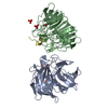
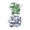
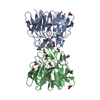
- PDB-4l7b: Structure of keap1 kelch domain with (1S,2R)-2-{[(1S)-1-[(1,3-dio... -
+ Open data

- Basic information
| Entry | Database: PDB / ID: 4l7b | ||||||
|---|---|---|---|---|---|---|---|
| Title | Structure of keap1 kelch domain with (1S,2R)-2-{[(1S)-1-[(1,3-dioxo-1,3-dihydro-2H-isoindol-2-yl)methyl]-3,4-dihydroisoquinolin-2(1H)-yl]carbonyl}cyclohexanecarboxylic acid | ||||||
 Components Components | Kelch-like ECH-associated protein 1 | ||||||
 Keywords Keywords | TRANSCRIPTION/INHIBITOR / STRESS SENSOR / KELCH DOMAIN / KELCH REPEAT MOTIF / BETA-PROPELLER / NRF2 / PROTEIN-SMALL MOLECULE COMPLEX / TRANSCRIPTION-INHIBITOR complex | ||||||
| Function / homology |  Function and homology information Function and homology informationregulation of epidermal cell differentiation / Nuclear events mediated by NFE2L2 / negative regulation of response to oxidative stress / Cul3-RING ubiquitin ligase complex / transcription regulator inhibitor activity / ubiquitin-like ligase-substrate adaptor activity / inclusion body / cellular response to interleukin-4 / regulation of autophagy / actin filament ...regulation of epidermal cell differentiation / Nuclear events mediated by NFE2L2 / negative regulation of response to oxidative stress / Cul3-RING ubiquitin ligase complex / transcription regulator inhibitor activity / ubiquitin-like ligase-substrate adaptor activity / inclusion body / cellular response to interleukin-4 / regulation of autophagy / actin filament / centriolar satellite / disordered domain specific binding / KEAP1-NFE2L2 pathway / positive regulation of proteasomal ubiquitin-dependent protein catabolic process / Antigen processing: Ubiquitination & Proteasome degradation / Neddylation / cellular response to oxidative stress / midbody / Potential therapeutics for SARS / in utero embryonic development / ubiquitin-dependent protein catabolic process / RNA polymerase II-specific DNA-binding transcription factor binding / proteasome-mediated ubiquitin-dependent protein catabolic process / Ub-specific processing proteases / protein ubiquitination / negative regulation of transcription by RNA polymerase II / endoplasmic reticulum / nucleoplasm / identical protein binding / cytoplasm / cytosol Similarity search - Function | ||||||
| Biological species |  Homo sapiens (human) Homo sapiens (human) | ||||||
| Method |  X-RAY DIFFRACTION / SYNCHROTRON / MOLECULAR REPLACEMENT / Resolution: 2.41 Å X-RAY DIFFRACTION / SYNCHROTRON / MOLECULAR REPLACEMENT / Resolution: 2.41 Å | ||||||
 Authors Authors | Jnoff, E. / Brookfield, F. / Albrecht, C. / Barker, J.J. / Barker, O. / Beaumont, E. / Bromidge, S. / Brooks, M. / Ceska, T. / Courade, J.P. ...Jnoff, E. / Brookfield, F. / Albrecht, C. / Barker, J.J. / Barker, O. / Beaumont, E. / Bromidge, S. / Brooks, M. / Ceska, T. / Courade, J.P. / Crabbe, T. / Duclos, S. / Fryatt, T. / Jigorel, E. / Kwong, J. / Sands, Z. / Smith, M.A. | ||||||
 Citation Citation | Journal: Chemmedchem / Year: 2014 Title: Binding Mode and Structure-Activity Relationships around Direct Inhibitors of the Nrf2-Keap1 Complex. Authors: Jnoff, E. / Albrecht, C. / Barker, J.J. / Barker, O. / Beaumont, E. / Bromidge, S. / Brookfield, F. / Brooks, M. / Bubert, C. / Ceska, T. / Corden, V. / Dawson, G. / Duclos, S. / Fryatt, T. ...Authors: Jnoff, E. / Albrecht, C. / Barker, J.J. / Barker, O. / Beaumont, E. / Bromidge, S. / Brookfield, F. / Brooks, M. / Bubert, C. / Ceska, T. / Corden, V. / Dawson, G. / Duclos, S. / Fryatt, T. / Genicot, C. / Jigorel, E. / Kwong, J. / Maghames, R. / Mushi, I. / Pike, R. / Sands, Z.A. / Smith, M.A. / Stimson, C.C. / Courade, J.P. | ||||||
| History |
|
- Structure visualization
| Structure viewer | Molecule: MolmilJmol/JSmol |
|---|
- Downloads & links
-Download
| PDBx/mmCIF format | 4l7b.cif.gz | 129.9 KB | Display | PDBx/mmCIF format |
|---|---|---|---|---|
| PDB format | pdb4l7b.ent.gz | 99.6 KB | Display | PDB format |
| PDBx/mmJSON format | 4l7b.json.gz | Tree view | PDBx/mmJSON format | |
| Others |  Other downloads Other downloads |
-Validation report
| Arichive directory | https://data.pdbj.org/pub/pdb/validation_reports/l7/4l7bftp://data.pdbj.org/pub/pdb/validation_reports/l7/4l7b | HTTPS FTP |
|---|
-Related structure data
| Related structure data | 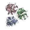 4l7cC 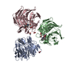 4l7dC 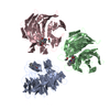 4n1bC  1zgkS C: citing same article ( S: Starting model for refinement |
|---|---|
| Similar structure data |


-Links
 PDBj
PDBj


- Assembly
| Deposited unit | 
| ||||||||
|---|---|---|---|---|---|---|---|---|---|
| 1 |
| ||||||||
| Unit cell |
|
-Components
| #1: Protein | Mass: 33009.801 Da / Num. of mol.: 2 / Fragment: Kelch domain, UNP residues 321-609 / Mutation: R354D Source method: isolated from a genetically manipulated source Source: (gene. exp.) Homo sapiens (human) / Gene: KEAP1, INRF2, KIAA0132, KLHL19 / Plasmid: pET28a / Production host:  #2: Chemical | ChemComp-ACT /   Mass: 59.044 Da / Num. of mol.: 5 / Source method: obtained synthetically / Formula: C2H3O2 Mass: 59.044 Da / Num. of mol.: 5 / Source method: obtained synthetically / Formula: C2H3O2#3: Chemical | ChemComp-1VV / ( |   Mass: 446.495 Da / Num. of mol.: 1 / Source method: obtained synthetically / Formula: C26H26N2O5 Mass: 446.495 Da / Num. of mol.: 1 / Source method: obtained synthetically / Formula: C26H26N2O5#4: Chemical | ChemComp-NA / |   Mass: 22.990 Da / Num. of mol.: 1 / Source method: obtained synthetically / Formula: Na Mass: 22.990 Da / Num. of mol.: 1 / Source method: obtained synthetically / Formula: Na#5: Water | ChemComp-HOH / |  Mass: 18.015 Da / Num. of mol.: 238 / Source method: isolated from a natural source / Formula: H2O Mass: 18.015 Da / Num. of mol.: 238 / Source method: isolated from a natural source / Formula: H2OHas protein modification | Y | |
|---|
-Experimental details
-Experiment
| Experiment | Method: X-RAY DIFFRACTION / Number of used crystals: 1 |
|---|
- Sample preparation
| Crystal | Density Matthews: 4.47 Å3/Da / Density % sol: 72.48 % |
|---|---|
| Crystal grow | Temperature: 296 K / Method: vapor diffusion, hanging drop / pH: 4.6 Details: 1 ul of protein at 12 mg/ml in 20 mM Tris-HCl pH 7.5, 5 mM DTT, 5 mM compound 1 + 2 ul of 2.15 M Sodium Acetate , VAPOR DIFFUSION, HANGING DROP, temperature 296K |
-Data collection
| Diffraction | Mean temperature: 100 K |
|---|---|
| Diffraction source | Source: SYNCHROTRON / Site: Diamond  / Beamline: I02 / Wavelength: 0.9795 Å / Beamline: I02 / Wavelength: 0.9795 Å |
| Detector | Type: DECTRIS PILATUS 6M / Detector: PIXEL / Date: Sep 19, 2012 / Details: Mirrors |
| Radiation | Monochromator: Double Crystal Monochromator / Protocol: SINGLE WAVELENGTH / Monochromatic (M) / Laue (L): M / Scattering type: x-ray |
| Radiation wavelength | Wavelength: 0.9795 Å / Relative weight: 1 |
| Reflection | Resolution: 2.41→75.37 Å / Num. all: 46220 / Num. obs: 46220 / % possible obs: 99.6 % / Observed criterion σ(F): 2 / Observed criterion σ(I): 2 / Biso Wilson estimate: 62.774 Å2 / Rmerge(I) obs: 0.064 |
| Reflection shell | Resolution: 2.41→2.47 Å / % possible all: 99.71 |
- Processing
| Software |
| ||||||||||||||||||||||||||||||||||||||||||||||||||||||||||||||||||||||||||||||||||||||||||||||||||||||||||||||||||||||||||||||||||||||||||||||||||||||||||||||||||||||||||
|---|---|---|---|---|---|---|---|---|---|---|---|---|---|---|---|---|---|---|---|---|---|---|---|---|---|---|---|---|---|---|---|---|---|---|---|---|---|---|---|---|---|---|---|---|---|---|---|---|---|---|---|---|---|---|---|---|---|---|---|---|---|---|---|---|---|---|---|---|---|---|---|---|---|---|---|---|---|---|---|---|---|---|---|---|---|---|---|---|---|---|---|---|---|---|---|---|---|---|---|---|---|---|---|---|---|---|---|---|---|---|---|---|---|---|---|---|---|---|---|---|---|---|---|---|---|---|---|---|---|---|---|---|---|---|---|---|---|---|---|---|---|---|---|---|---|---|---|---|---|---|---|---|---|---|---|---|---|---|---|---|---|---|---|---|---|---|---|---|---|---|---|
| Refinement | Method to determine structure: MOLECULAR REPLACEMENT Starting model: PDB ENTRY 1ZGK Resolution: 2.41→60.96 Å / Cor.coef. Fo:Fc: 0.937 / Cor.coef. Fo:Fc free: 0.927 / SU B: 7.271 / SU ML: 0.161 / Cross valid method: THROUGHOUT / ESU R: 0.247 / ESU R Free: 0.2 / Stereochemistry target values: MAXIMUM LIKELIHOOD / Details: HYDROGENS HAVE BEEN ADDED IN THE RIDING POSITIONS
| ||||||||||||||||||||||||||||||||||||||||||||||||||||||||||||||||||||||||||||||||||||||||||||||||||||||||||||||||||||||||||||||||||||||||||||||||||||||||||||||||||||||||||
| Solvent computation | Ion probe radii: 0.8 Å / Shrinkage radii: 0.8 Å / VDW probe radii: 1.2 Å / Solvent model: MASK | ||||||||||||||||||||||||||||||||||||||||||||||||||||||||||||||||||||||||||||||||||||||||||||||||||||||||||||||||||||||||||||||||||||||||||||||||||||||||||||||||||||||||||
| Displacement parameters | Biso mean: 51.371 Å2
| ||||||||||||||||||||||||||||||||||||||||||||||||||||||||||||||||||||||||||||||||||||||||||||||||||||||||||||||||||||||||||||||||||||||||||||||||||||||||||||||||||||||||||
| Refinement step | Cycle: LAST / Resolution: 2.41→60.96 Å
| ||||||||||||||||||||||||||||||||||||||||||||||||||||||||||||||||||||||||||||||||||||||||||||||||||||||||||||||||||||||||||||||||||||||||||||||||||||||||||||||||||||||||||
| Refine LS restraints |
| ||||||||||||||||||||||||||||||||||||||||||||||||||||||||||||||||||||||||||||||||||||||||||||||||||||||||||||||||||||||||||||||||||||||||||||||||||||||||||||||||||||||||||
| LS refinement shell | Resolution: 2.41→2.473 Å / Total num. of bins used: 20
|
