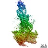
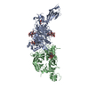
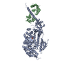
Crystal Structure of nucleotide-free Myosin 1b residues 1-728 with bound Calmodulin
要素
Calmodulin
Unconventional myosin-Ib
キーワード
MOTOR PROTEIN/METAL BINDING PROTEIN / myosin motor / actin binding / nucleotide hydrolysis / cargo / membrane binding / ca2+ binding / MOTOR PROTEIN-METAL BINDING PROTEIN complex
機能・相同性
機能・相同性情報
post-Golgi vesicle-mediated transport / : / : / : / actin filament-based movement / : / : / positive regulation of protein autophosphorylation / negative regulation of peptidyl-threonine phosphorylation / establishment of protein localization to mitochondrial membrane ...post-Golgi vesicle-mediated transport / : / : / : / actin filament-based movement / : / : / positive regulation of protein autophosphorylation / negative regulation of peptidyl-threonine phosphorylation / establishment of protein localization to mitochondrial membrane / type 3 metabotropic glutamate receptor binding / myosin complex / CaM pathway / positive regulation of peptidyl-threonine phosphorylation / Cam-PDE 1 activation / Sodium/Calcium exchangers / Calmodulin induced events / positive regulation of DNA binding / Reduction of cytosolic Ca++ levels / Activation of Ca-permeable Kainate Receptor / CREB1 phosphorylation through the activation of CaMKII/CaMKK/CaMKIV cascasde / Loss of phosphorylation of MECP2 at T308 / CREB1 phosphorylation through the activation of Adenylate Cyclase / response to corticosterone / CaMK IV-mediated phosphorylation of CREB / PKA activation / negative regulation of high voltage-gated calcium channel activity / Glycogen breakdown (glycogenolysis) / CLEC7A (Dectin-1) induces NFAT activation / Activation of RAC1 downstream of NMDARs / negative regulation of ryanodine-sensitive calcium-release channel activity / organelle localization by membrane tethering / microfilament motor activity / mitochondrion-endoplasmic reticulum membrane tethering / autophagosome membrane docking / negative regulation of calcium ion export across plasma membrane / regulation of cardiac muscle cell action potential / presynaptic endocytosis / nitric-oxide synthase binding / Synthesis of IP3 and IP4 in the cytosol / regulation of synaptic vesicle exocytosis / regulation of cell communication by electrical coupling involved in cardiac conduction / microvillus / Phase 0 - rapid depolarisation / calcineurin-mediated signaling / Negative regulation of NMDA receptor-mediated neuronal transmission / Unblocking of NMDA receptors, glutamate binding and activation / phosphatidylinositol-3,4,5-trisphosphate binding / RHO GTPases activate PAKs / Ion transport by P-type ATPases / brush border / Uptake and function of anthrax toxins / cytoskeletal motor activity / actin filament bundle assembly / adenylate cyclase binding / regulation of ryanodine-sensitive calcium-release channel activity / protein phosphatase activator activity / Long-term potentiation / Calcineurin activates NFAT / Regulation of MECP2 expression and activity / positive regulation of protein serine/threonine kinase activity / DARPP-32 events / catalytic complex / Smooth Muscle Contraction / detection of calcium ion / regulation of synaptic vesicle endocytosis / regulation of cardiac muscle contraction / RHO GTPases activate IQGAPs / regulation of cardiac muscle contraction by regulation of the release of sequestered calcium ion / activation of adenylate cyclase activity / cellular response to interferon-beta / Protein methylation / phosphatidylinositol 3-kinase binding / calcium channel inhibitor activity / Activation of AMPK downstream of NMDARs / presynaptic cytosol / positive regulation of nitric-oxide synthase activity / Ion homeostasis / regulation of release of sequestered calcium ion into cytosol by sarcoplasmic reticulum / enzyme regulator activity / eNOS activation / titin binding / Tetrahydrobiopterin (BH4) synthesis, recycling, salvage and regulation / sperm midpiece / regulation of calcium-mediated signaling / phosphatidylinositol-4,5-bisphosphate binding / voltage-gated potassium channel complex / calcium channel complex / FCERI mediated Ca+2 mobilization / substantia nigra development / Ras activation upon Ca2+ influx through NMDA receptor / regulation of heart rate / FCGR3A-mediated IL10 synthesis / Antigen activates B Cell Receptor (BCR) leading to generation of second messengers / calyx of Held / actin filament organization / response to amphetamine / adenylate cyclase activator activity / sarcomere / VEGFR2 mediated cell proliferation 類似検索 - 分子機能
Single alpha-helices involved in coiled-coils or other helix-helix interfaces - #4820 / Class I myosin tail homology domain / Class I myosin, motor domain / Unconventional myosin tail, actin- and lipid-binding / Class I myosin tail homology (TH1) domain profile. / IQ calmodulin-binding motif / Short calmodulin-binding motif containing conserved Ile and Gln residues. / IQ motif, EF-hand binding site / Myosin head, motor domain / Myosin head (motor domain) ...Single alpha-helices involved in coiled-coils or other helix-helix interfaces - #4820 / Class I myosin tail homology domain / Class I myosin, motor domain / Unconventional myosin tail, actin- and lipid-binding / Class I myosin tail homology (TH1) domain profile. / IQ calmodulin-binding motif / Short calmodulin-binding motif containing conserved Ile and Gln residues. / IQ motif, EF-hand binding site / Myosin head, motor domain / Myosin head (motor domain) / Myosin motor domain profile. / Myosin. Large ATPases. / IQ motif profile. / Kinesin motor domain superfamily / : / Single alpha-helices involved in coiled-coils or other helix-helix interfaces / EF-hand domain pair / EF-hand, calcium binding motif / EF-Hand 1, calcium-binding site / EF-hand calcium-binding domain. / EF-hand calcium-binding domain profile. / EF-hand domain / EF-hand domain pair / Up-down Bundle / P-loop containing nucleoside triphosphate hydrolase / Mainly Alpha 類似検索 - ドメイン・相同性
 ムービー
ムービー コントローラー


 データを開く
 基本情報
基本情報 要素
要素 キーワード
キーワード 機能・相同性情報
 Homo sapiens (ヒト)
 X線回折 /
  データ登録者
 引用
引用 構造の表示
構造の表示 ダウンロードとリンク
 その他のダウンロード
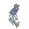


 PDBj
PDBj


 集合体
集合体

 Spodoptera frugiperda (ツマジロクサヨトウ)
 分子量: 24.305 Da / 分子数: 1 / 由来タイプ: 合成 / 式: Mg
 分子量: 18.015 Da / 分子数: 459 / 由来タイプ: 天然 / 式: H2O
 試料調製
試料調製 / ビームライン: X6A / 波長: 1 Å
 解析
解析