 Movie
Movie Controller

[English] 日本語
 Yorodumi
- PDB-4l79: Crystal Structure of nucleotide-free Myosin 1b residues 1-728 wit... -
+ Open data

- Basic information
| Entry | Database: PDB / ID: 4l79 | ||||||
|---|---|---|---|---|---|---|---|
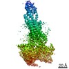
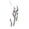
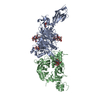
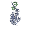
| Title | Crystal Structure of nucleotide-free Myosin 1b residues 1-728 with bound Calmodulin | ||||||
 Components Components |
| ||||||
 Keywords Keywords | MOTOR PROTEIN/METAL BINDING PROTEIN / myosin motor / actin binding / nucleotide hydrolysis / cargo / membrane binding / ca2+ binding / MOTOR PROTEIN-METAL BINDING PROTEIN complex | ||||||
| Function / homology |  Function and homology information Function and homology informationpost-Golgi vesicle-mediated transport / : / : / : / : / positive regulation of protein autophosphorylation / : / negative regulation of peptidyl-threonine phosphorylation / actin filament-based movement / : ...post-Golgi vesicle-mediated transport / : / : / : / : / positive regulation of protein autophosphorylation / : / negative regulation of peptidyl-threonine phosphorylation / actin filament-based movement / : / type 3 metabotropic glutamate receptor binding / myosin complex / positive regulation of peptidyl-threonine phosphorylation / positive regulation of DNA binding / CaM pathway / Cam-PDE 1 activation / Sodium/Calcium exchangers / Calmodulin induced events / Reduction of cytosolic Ca++ levels / Activation of Ca-permeable Kainate Receptor / CREB1 phosphorylation through the activation of CaMKII/CaMKK/CaMKIV cascasde / Loss of phosphorylation of MECP2 at T308 / CREB1 phosphorylation through the activation of Adenylate Cyclase / negative regulation of high voltage-gated calcium channel activity / PKA activation / CaMK IV-mediated phosphorylation of CREB / Glycogen breakdown (glycogenolysis) / CLEC7A (Dectin-1) induces NFAT activation / microfilament motor activity / Activation of RAC1 downstream of NMDARs / negative regulation of ryanodine-sensitive calcium-release channel activity / organelle localization by membrane tethering / response to corticosterone / mitochondrion-endoplasmic reticulum membrane tethering / autophagosome membrane docking / negative regulation of calcium ion export across plasma membrane / regulation of synaptic vesicle exocytosis / regulation of ryanodine-sensitive calcium-release channel activity / regulation of cardiac muscle cell action potential / presynaptic endocytosis / Synthesis of IP3 and IP4 in the cytosol / Phase 0 - rapid depolarisation / phosphatidylinositol-3,4,5-trisphosphate binding / positive regulation of protein serine/threonine kinase activity / microvillus / Negative regulation of NMDA receptor-mediated neuronal transmission / Unblocking of NMDA receptors, glutamate binding and activation / RHO GTPases activate PAKs / calcineurin-mediated signaling / cytoskeletal motor activity / regulation of cell communication by electrical coupling involved in cardiac conduction / nitric-oxide synthase binding / Ion transport by P-type ATPases / brush border / actin filament bundle assembly / Uptake and function of anthrax toxins / adenylate cyclase binding / protein phosphatase activator activity / Long-term potentiation / Calcineurin activates NFAT / Regulation of MECP2 expression and activity / DARPP-32 events / Smooth Muscle Contraction / regulation of synaptic vesicle endocytosis / detection of calcium ion / regulation of cardiac muscle contraction / catalytic complex / RHO GTPases activate IQGAPs / phosphatidylinositol 3-kinase binding / positive regulation of nitric-oxide synthase activity / activation of adenylate cyclase activity / calcium channel inhibitor activity / presynaptic cytosol / cellular response to interferon-beta / Activation of AMPK downstream of NMDARs / enzyme regulator activity / Ion homeostasis / regulation of release of sequestered calcium ion into cytosol by sarcoplasmic reticulum / eNOS activation / Protein methylation / Tetrahydrobiopterin (BH4) synthesis, recycling, salvage and regulation / titin binding / regulation of calcium-mediated signaling / phosphatidylinositol-4,5-bisphosphate binding / regulation of cardiac muscle contraction by regulation of the release of sequestered calcium ion / voltage-gated potassium channel complex / FCERI mediated Ca+2 mobilization / calcium channel complex / substantia nigra development / FCGR3A-mediated IL10 synthesis / regulation of heart rate / Antigen activates B Cell Receptor (BCR) leading to generation of second messengers / Ras activation upon Ca2+ influx through NMDA receptor / calyx of Held / nitric-oxide synthase regulator activity / adenylate cyclase activator activity / trans-Golgi network membrane / VEGFR2 mediated cell proliferation / VEGFR2 mediated vascular permeability / regulation of cytokinesis Similarity search - Function | ||||||
| Biological species |   Homo sapiens (human) Homo sapiens (human) | ||||||
| Method |  X-RAY DIFFRACTION / SYNCHROTRON / MOLECULAR REPLACEMENT / Resolution: 2.3 Å X-RAY DIFFRACTION / SYNCHROTRON / MOLECULAR REPLACEMENT / Resolution: 2.3 Å | ||||||
 Authors Authors | Shuman, H. / Zwolak, A. / Dominguez, R. / Ostap, E.M. | ||||||
 Citation Citation | Journal: Proc.Natl.Acad.Sci.USA / Year: 2014 Title: A vertebrate myosin-I structure reveals unique insights into myosin mechanochemical tuning. Authors: Shuman, H. / Greenberg, M.J. / Zwolak, A. / Lin, T. / Sindelar, C.V. / Dominguez, R. / Ostap, E.M. | ||||||
| History |
|
- Structure visualization
| Structure viewer | Molecule: MolmilJmol/JSmol |
|---|
- Downloads & links
-Download
| PDBx/mmCIF format | 4l79.cif.gz | 200.4 KB | Display | PDBx/mmCIF format |
|---|---|---|---|---|
| PDB format | pdb4l79.ent.gz | 158 KB | Display | PDB format |
| PDBx/mmJSON format | 4l79.json.gz | Tree view | PDBx/mmJSON format | |
| Others |  Other downloads Other downloads |
-Validation report
| Arichive directory | https://data.pdbj.org/pub/pdb/validation_reports/l7/4l79ftp://data.pdbj.org/pub/pdb/validation_reports/l7/4l79 | HTTPS FTP |
|---|
-Related structure data
| Related structure data | 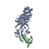 1oe9S S: Starting model for refinement |
|---|---|
| Similar structure data |


-Links
 PDBj
PDBj


- Assembly
| Deposited unit | 
| ||||||||
|---|---|---|---|---|---|---|---|---|---|
| 1 |
| ||||||||
| Unit cell |
|
-Components
| #1: Protein | Mass: 86002.008 Da / Num. of mol.: 1 / Fragment: UNP residues 1-728 Source method: isolated from a genetically manipulated source Source: (gene. exp.)   Spodoptera frugiperda (fall armyworm) / References: UniProt: Q05096 Spodoptera frugiperda (fall armyworm) / References: UniProt: Q05096 |
|---|---|
| #2: Protein | Mass: 16852.545 Da / Num. of mol.: 1 Source method: isolated from a genetically manipulated source Source: (gene. exp.) Homo sapiens (human)Gene: CALM, CALM1, CALM2, CALM3, CALML2, calmodulin, CAM, CAM1, CAM2, CAM3, CAMB, CAMC, CAMIII Production host: Spodoptera frugiperda (fall armyworm) / References: UniProt: P62158, UniProt: P0DP23*PLUS |
| #3: Chemical | ChemComp-MG /   Mass: 24.305 Da / Num. of mol.: 1 / Source method: obtained synthetically / Formula: Mg Mass: 24.305 Da / Num. of mol.: 1 / Source method: obtained synthetically / Formula: Mg |
| #4: Water | ChemComp-HOH /  Mass: 18.015 Da / Num. of mol.: 459 / Source method: isolated from a natural source / Formula: H2O Mass: 18.015 Da / Num. of mol.: 459 / Source method: isolated from a natural source / Formula: H2O |
-Experimental details
-Experiment
| Experiment | Method: X-RAY DIFFRACTION / Number of used crystals: 1 |
|---|
- Sample preparation
| Crystal | Density Matthews: 2.57 Å3/Da / Density % sol: 52.19 % |
|---|---|
| Crystal grow | Temperature: 277 K / Method: vapor diffusion, hanging drop / pH: 7 Details: 10% PEG 3350, 100 mM LiOOCCH3, 100 mM HEPES, pH 7.0, VAPOR DIFFUSION, HANGING DROP, temperature 277K |
-Data collection
| Diffraction | Mean temperature: 100 K |
|---|---|
| Diffraction source | Source: SYNCHROTRON / Site: NSLS  / Beamline: X6A / Wavelength: 1 Å / Beamline: X6A / Wavelength: 1 Å |
| Detector | Type: ADSC QUANTUM 270 / Detector: CCD / Date: Oct 10, 2012 / Details: TOROIDAL FOCUSING MIRROR |
| Radiation | Monochromator: SI(111) CHANNEL CUT / Protocol: SINGLE WAVELENGTH / Monochromatic (M) / Laue (L): M / Scattering type: x-ray |
| Radiation wavelength | Wavelength: 1 Å / Relative weight: 1 |
| Reflection | Resolution: 2.3→40 Å / Num. obs: 52157 / % possible obs: 99 % / Observed criterion σ(I): 2 |
| Reflection shell | Resolution: 2.3→2.4 Å / % possible all: 87.8 |
- Processing
| Software |
| ||||||||||||||||||||||||||||||||||||||||||||||||||||||||||||||||||||||||||||||||||||||||||||||||||||||||||||||||||||||||||||||
|---|---|---|---|---|---|---|---|---|---|---|---|---|---|---|---|---|---|---|---|---|---|---|---|---|---|---|---|---|---|---|---|---|---|---|---|---|---|---|---|---|---|---|---|---|---|---|---|---|---|---|---|---|---|---|---|---|---|---|---|---|---|---|---|---|---|---|---|---|---|---|---|---|---|---|---|---|---|---|---|---|---|---|---|---|---|---|---|---|---|---|---|---|---|---|---|---|---|---|---|---|---|---|---|---|---|---|---|---|---|---|---|---|---|---|---|---|---|---|---|---|---|---|---|---|---|---|---|
| Refinement | Method to determine structure: MOLECULAR REPLACEMENT Starting model: PDB entry 1oe9 Resolution: 2.3→39.209 Å / SU ML: 0.28 / σ(F): 1.34 / Phase error: 25.01 / Stereochemistry target values: ML
| ||||||||||||||||||||||||||||||||||||||||||||||||||||||||||||||||||||||||||||||||||||||||||||||||||||||||||||||||||||||||||||||
| Solvent computation | Shrinkage radii: 0.9 Å / VDW probe radii: 1.11 Å / Solvent model: FLAT BULK SOLVENT MODEL | ||||||||||||||||||||||||||||||||||||||||||||||||||||||||||||||||||||||||||||||||||||||||||||||||||||||||||||||||||||||||||||||
| Refinement step | Cycle: LAST / Resolution: 2.3→39.209 Å
| ||||||||||||||||||||||||||||||||||||||||||||||||||||||||||||||||||||||||||||||||||||||||||||||||||||||||||||||||||||||||||||||
| Refine LS restraints |
| ||||||||||||||||||||||||||||||||||||||||||||||||||||||||||||||||||||||||||||||||||||||||||||||||||||||||||||||||||||||||||||||
| LS refinement shell |
|
