 Movie
Movie Controller

[English] 日本語
 Yorodumi
- PDB-4l1i: Crystal structure of EGFP-based Calcium Sensor CatchER complexed ... -
+ Open data

- Basic information
| Entry | Database: PDB / ID: 4l1i | ||||||
|---|---|---|---|---|---|---|---|
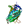
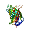
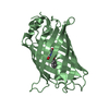
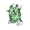
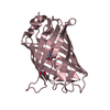
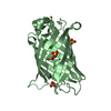
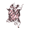
| Title | Crystal structure of EGFP-based Calcium Sensor CatchER complexed with Ca | ||||||
 Components Components | EGFP-based Calcium Sensor CatchER | ||||||
 Keywords Keywords | CALCIUM BINDING PROTEIN / chromophore / luminescence / fluorescent protein / calcium sensor / metal ions / gadolinium | ||||||
| Function / homology |  Function and homology information Function and homology information | ||||||
| Biological species |   Aequorea victoria (jellyfish) Aequorea victoria (jellyfish) | ||||||
| Method |  X-RAY DIFFRACTION / SYNCHROTRON / MOLECULAR REPLACEMENT / Resolution: 1.2 Å X-RAY DIFFRACTION / SYNCHROTRON / MOLECULAR REPLACEMENT / Resolution: 1.2 Å | ||||||
 Authors Authors | Zhang, Y. / Weber, I.T. | ||||||
 Citation Citation | Journal: Acta Crystallogr.,Sect.D / Year: 2013 Title: Structural basis for a hand-like site in the calcium sensor CatchER with fast kinetics. Authors: Zhang, Y. / Reddish, F. / Tang, S. / Zhuo, Y. / Wang, Y.F. / Yang, J.J. / Weber, I.T. | ||||||
| History |
|
- Structure visualization
| Structure viewer | Molecule: MolmilJmol/JSmol |
|---|
- Downloads & links
-Download
| PDBx/mmCIF format | 4l1i.cif.gz | 119.6 KB | Display | PDBx/mmCIF format |
|---|---|---|---|---|
| PDB format | pdb4l1i.ent.gz | 91.6 KB | Display | PDB format |
| PDBx/mmJSON format | 4l1i.json.gz | Tree view | PDBx/mmJSON format | |
| Others |  Other downloads Other downloads |
-Validation report
| Summary document | 4l1i_validation.pdf.gz | 433 KB | Display | wwPDB validaton report |
|---|---|---|---|---|
| Full document | 4l1i_full_validation.pdf.gz | 436.8 KB | Display | |
| Data in XML | 4l1i_validation.xml.gz | 13.5 KB | Display | |
| Data in CIF | 4l1i_validation.cif.gz | 19.5 KB | Display | |
| Arichive directory | https://data.pdbj.org/pub/pdb/validation_reports/l1/4l1iftp://data.pdbj.org/pub/pdb/validation_reports/l1/4l1i | HTTPS FTP |
-Related structure data
| Related structure data |  4l12C  4l13C  2okwS C: citing same article ( S: Starting model for refinement |
|---|---|
| Similar structure data |


-Links
 PDBj
PDBj

- Assembly
| Deposited unit | 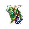
| ||||||||
|---|---|---|---|---|---|---|---|---|---|
| 1 |
| ||||||||
| Unit cell |
|
-Components
| #1: Protein | Mass: 26141.316 Da / Num. of mol.: 1 / Fragment: SEE REMARK 999 Mutation: F64L, S65T, S147E, M153T, V163A, S202D, Q204E, F223E, T225E Source method: isolated from a genetically manipulated source Source: (gene. exp.) Aequorea victoria (jellyfish) / Gene: GFP / Plasmid: pET28a / Production host:  | ||||||
|---|---|---|---|---|---|---|---|
| #2: Chemical |   Mass: 40.078 Da / Num. of mol.: 2 / Source method: obtained synthetically / Formula: Ca Mass: 40.078 Da / Num. of mol.: 2 / Source method: obtained synthetically / Formula: Ca#3: Water | ChemComp-HOH / |  Mass: 18.015 Da / Num. of mol.: 197 / Source method: isolated from a natural source / Formula: H2O Mass: 18.015 Da / Num. of mol.: 197 / Source method: isolated from a natural source / Formula: H2OHas protein modification | Y | Sequence details | RESIDUE SER 65 UNDERWENT MUTATION TO THR 65. THR 65, TYR 66, AND GLY 67 CIRCULARIZ | |
-Experimental details
-Experiment
| Experiment | Method: X-RAY DIFFRACTION / Number of used crystals: 1 |
|---|
- Sample preparation
| Crystal | Density Matthews: 2.13 Å3/Da / Density % sol: 42.37 % |
|---|---|
| Crystal grow | Temperature: 298 K / Method: vapor diffusion, hanging drop / pH: 7 Details: 0.9 mM protein, 53 mM HEPES, pH 7.0, 1 mM beta-mercaptoethanol, 50 mM sodium acetate, 16% PEG3350, VAPOR DIFFUSION, HANGING DROP, temperature 298K |
-Data collection
| Diffraction | Mean temperature: 100 K |
|---|---|
| Diffraction source | Source: SYNCHROTRON / Site: APS  / Beamline: 22-ID / Wavelength: 0.8 / Wavelength: 0.8 Å / Beamline: 22-ID / Wavelength: 0.8 / Wavelength: 0.8 Å |
| Detector | Type: MARMOSAIC 300 mm CCD / Detector: CCD / Date: Nov 17, 2010 |
| Radiation | Monochromator: double crystal Si(111) / Protocol: SINGLE WAVELENGTH / Monochromatic (M) / Laue (L): M / Scattering type: x-ray |
| Radiation wavelength | Wavelength: 0.8 Å / Relative weight: 1 |
| Reflection | Resolution: 1.2→50 Å / Num. all: 70349 / Num. obs: 70349 / % possible obs: 99.2 % / Observed criterion σ(F): 0 / Observed criterion σ(I): 0 / Redundancy: 12.5 % / Rmerge(I) obs: 0.081 / Net I/σ(I): 19.9 |
| Reflection shell | Resolution: 1.2→1.24 Å / Redundancy: 7.8 % / Rmerge(I) obs: 0.392 / Mean I/σ(I) obs: 8.7 / % possible all: 100 |
- Processing
| Software |
| |||||||||||||||||||||||||||||||||
|---|---|---|---|---|---|---|---|---|---|---|---|---|---|---|---|---|---|---|---|---|---|---|---|---|---|---|---|---|---|---|---|---|---|---|
| Refinement | Method to determine structure: MOLECULAR REPLACEMENT Starting model: PDB ENTRY 2OKW Resolution: 1.2→50 Å / Num. parameters: 19518 / Num. restraintsaints: 25624 / Cross valid method: FREE R / σ(F): 0 / Stereochemistry target values: ENGH AND HUBER Details: ANISOTROPIC REFINEMENT REDUCED FREE R (NO CUTOFF) BY ?
| |||||||||||||||||||||||||||||||||
| Refine analyze | Num. disordered residues: 23 / Occupancy sum hydrogen: 0 / Occupancy sum non hydrogen: 2019.61 | |||||||||||||||||||||||||||||||||
| Refinement step | Cycle: LAST / Resolution: 1.2→50 Å
| |||||||||||||||||||||||||||||||||
| Refine LS restraints |
|
