 Movie
Movie Controller


+ Open data

- Basic information
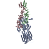
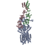
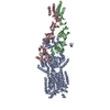
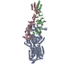
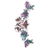
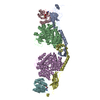
| Entry | Database: PDB / ID: 4ktt | ||||||
|---|---|---|---|---|---|---|---|
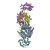
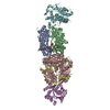
| Title | Structural insights of MAT enzymes: MATa2b complexed with SAM | ||||||
 Components Components |
| ||||||
 Keywords Keywords | TRANSFERASE / SAMe Synthesis | ||||||
| Function / homology |  Function and homology information Function and homology informationmethionine adenosyltransferase regulator activity / methionine adenosyltransferase complex / methionine adenosyltransferase / methionine adenosyltransferase activity / protein heterooligomerization / S-adenosylmethionine biosynthetic process / Methylation / cellular response to methionine / protein hexamerization / small molecule binding ...methionine adenosyltransferase regulator activity / methionine adenosyltransferase complex / methionine adenosyltransferase / methionine adenosyltransferase activity / protein heterooligomerization / S-adenosylmethionine biosynthetic process / Methylation / cellular response to methionine / protein hexamerization / small molecule binding / enzyme regulator activity / one-carbon metabolic process / positive regulation of TORC1 signaling / Ub-specific processing proteases / enzyme binding / extracellular exosome / ATP binding / metal ion binding / identical protein binding / nucleus / cytosol Similarity search - Function | ||||||
| Biological species |  Homo sapiens (human) Homo sapiens (human) | ||||||
| Method |  X-RAY DIFFRACTION / SYNCHROTRON / MOLECULAR REPLACEMENT / Resolution: 2.59 Å X-RAY DIFFRACTION / SYNCHROTRON / MOLECULAR REPLACEMENT / Resolution: 2.59 Å | ||||||
 Authors Authors | Murray, B. / Antonyuk, S.V. / Marina, A. / Lu, S.C. / Mato, J.M. / Hasnain, S.S. / Rojas, A.L. | ||||||
 Citation Citation | Journal: IUCrJ / Year: 2014 Title: Structure and function study of the complex that synthesizes S-adenosylmethionine. Authors: Murray, B. / Antonyuk, S.V. / Marina, A. / Van Liempd, S.M. / Lu, S.C. / Mato, J.M. / Hasnain, S.S. / Rojas, A.L. | ||||||
| History |
|
- Structure visualization
| Structure viewer | Molecule: MolmilJmol/JSmol |
|---|
- Downloads & links
-Download
| PDBx/mmCIF format | 4ktt.cif.gz | 417.8 KB | Display | PDBx/mmCIF format |
|---|---|---|---|---|
| PDB format | pdb4ktt.ent.gz | 337 KB | Display | PDB format |
| PDBx/mmJSON format | 4ktt.json.gz | Tree view | PDBx/mmJSON format | |
| Others |  Other downloads Other downloads |
-Validation report
| Arichive directory | https://data.pdbj.org/pub/pdb/validation_reports/kt/4kttftp://data.pdbj.org/pub/pdb/validation_reports/kt/4ktt | HTTPS FTP |
|---|
-Related structure data
| Related structure data |  4ktvC  4ndnC 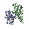 2po2S  2ydyS C: citing same article ( S: Starting model for refinement |
|---|---|
| Similar structure data |


-Links
 PDBj
PDBj
- Assembly
| Deposited unit | 
| ||||||||
|---|---|---|---|---|---|---|---|---|---|
| 1 |
| ||||||||
| Unit cell |
|
-Components
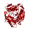
-Protein , 2 types, 6 molecules ABCDEF
| #1: Protein | Mass: 43807.703 Da / Num. of mol.: 4 Source method: isolated from a genetically manipulated source Source: (gene. exp.) Homo sapiens (human) / Gene: MAT2A, AMS2, MATA2 / Production host:  #2: Protein | Mass: 36862.820 Da / Num. of mol.: 2 Source method: isolated from a genetically manipulated source Source: (gene. exp.) Homo sapiens (human) / Gene: MAT2B, TGR, MSTP045, Nbla02999, UNQ2435/PRO4995 / Production host: |
|---|
-Non-polymers , 5 types, 141 molecules 


| #3: Chemical | ChemComp-EDO /  Mass: 62.068 Da / Num. of mol.: 28 / Source method: obtained synthetically / Formula: C2H6O2 Mass: 62.068 Da / Num. of mol.: 28 / Source method: obtained synthetically / Formula: C2H6O2#4: Chemical |  Mass: 398.437 Da / Num. of mol.: 2 / Source method: obtained synthetically / Formula: C15H22N6O5S Mass: 398.437 Da / Num. of mol.: 2 / Source method: obtained synthetically / Formula: C15H22N6O5S#5: Chemical |  Mass: 94.971 Da / Num. of mol.: 2 / Source method: obtained synthetically / Formula: PO4 Mass: 94.971 Da / Num. of mol.: 2 / Source method: obtained synthetically / Formula: PO4#6: Chemical | ChemComp-MG / |  Mass: 24.305 Da / Num. of mol.: 1 / Source method: obtained synthetically / Formula: Mg Mass: 24.305 Da / Num. of mol.: 1 / Source method: obtained synthetically / Formula: Mg#7: Water | ChemComp-HOH / | Mass: 18.015 Da / Num. of mol.: 108 / Source method: isolated from a natural source / Formula: H2O |
|---|
-Details
| Sequence details | THE DATABASE SEQEUNCE REFEREENCE FOR METHIONINE ADENOSYLTRANSFERASE 2 SUBUNIT BETA IS Q9NZL9 ...THE DATABASE SEQEUNCE REFEREENCE |
|---|
-Experimental details
-Experiment
| Experiment | Method: X-RAY DIFFRACTION / Number of used crystals: 1 |
|---|
- Sample preparation
| Crystal | Density Matthews: 2.51 Å3/Da / Density % sol: 51.04 % |
|---|---|
| Crystal grow | Temperature: 298 K / pH: 6.5 Details: 0.1M MES/Imidazole pH 6.5 0.1M Carboxylic acids 20% Ethylene glycol 10% PEG 8K, VAPOR DIFFUSION, HANGING DROP, temperature 298K |
-Data collection
| Diffraction | Mean temperature: 100 K |
|---|---|
| Diffraction source | Source: SYNCHROTRON / Site: SOLEIL  / Beamline: PROXIMA 1 / Wavelength: 0.98 / Beamline: PROXIMA 1 / Wavelength: 0.98 |
| Detector | Type: PSI PILATUS 6M / Detector: PIXEL / Date: Feb 20, 2013 |
| Radiation | Protocol: SINGLE WAVELENGTH / Monochromatic (M) / Laue (L): M / Scattering type: x-ray |
| Radiation wavelength | Wavelength: 0.98 Å / Relative weight: 1 |
| Reflection | Resolution: 2.59→50 Å / Num. all: 78380 / Num. obs: 77361 / % possible obs: 98.7 % / Observed criterion σ(F): 1.75 / Observed criterion σ(I): 1.75 / Redundancy: 5.6 % / Rmerge(I) obs: 0.144 |
| Reflection shell | Resolution: 2.6→2.69 Å / % possible all: 90.1 |
- Processing
| Software |
| ||||||||||||||||||||||||||||||||||||||||||||||||||||||||||||||||||||||||||||||||||||||||||||||||||||||||||||||||||||||||||||||||||||||||||||||||||||||||||||||||||||||||||||||||||||||
|---|---|---|---|---|---|---|---|---|---|---|---|---|---|---|---|---|---|---|---|---|---|---|---|---|---|---|---|---|---|---|---|---|---|---|---|---|---|---|---|---|---|---|---|---|---|---|---|---|---|---|---|---|---|---|---|---|---|---|---|---|---|---|---|---|---|---|---|---|---|---|---|---|---|---|---|---|---|---|---|---|---|---|---|---|---|---|---|---|---|---|---|---|---|---|---|---|---|---|---|---|---|---|---|---|---|---|---|---|---|---|---|---|---|---|---|---|---|---|---|---|---|---|---|---|---|---|---|---|---|---|---|---|---|---|---|---|---|---|---|---|---|---|---|---|---|---|---|---|---|---|---|---|---|---|---|---|---|---|---|---|---|---|---|---|---|---|---|---|---|---|---|---|---|---|---|---|---|---|---|---|---|---|---|
| Refinement | Method to determine structure: MOLECULAR REPLACEMENT Starting model: PDB ENTRIES 2PO2 AND 2YDY Resolution: 2.59→47.41 Å / Cor.coef. Fo:Fc: 0.945 / Cor.coef. Fo:Fc free: 0.909 / Occupancy max: 1 / Occupancy min: 0.52 / SU B: 12.967 / SU ML: 0.275 / Stereochemistry target values: ML / Details: HYDROGENS HAVE BEEN ADDED IN THE
| ||||||||||||||||||||||||||||||||||||||||||||||||||||||||||||||||||||||||||||||||||||||||||||||||||||||||||||||||||||||||||||||||||||||||||||||||||||||||||||||||||||||||||||||||||||||
| Solvent computation | Solvent model: MASK | ||||||||||||||||||||||||||||||||||||||||||||||||||||||||||||||||||||||||||||||||||||||||||||||||||||||||||||||||||||||||||||||||||||||||||||||||||||||||||||||||||||||||||||||||||||||
| Displacement parameters | Biso mean: 75.2 Å2
| ||||||||||||||||||||||||||||||||||||||||||||||||||||||||||||||||||||||||||||||||||||||||||||||||||||||||||||||||||||||||||||||||||||||||||||||||||||||||||||||||||||||||||||||||||||||
| Refinement step | Cycle: LAST / Resolution: 2.59→47.41 Å
| ||||||||||||||||||||||||||||||||||||||||||||||||||||||||||||||||||||||||||||||||||||||||||||||||||||||||||||||||||||||||||||||||||||||||||||||||||||||||||||||||||||||||||||||||||||||
| Refine LS restraints |
|