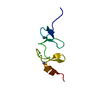
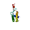
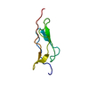
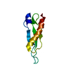
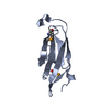
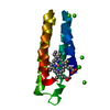
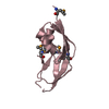
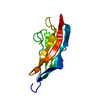
[English] 日本語
Yorodumi
- PDB-4kdw: Crystal structure of a bacterial immunoglobulin-like domain from ... -

+
Open data


ID or keywords:

Loading...

-
Basic information

Entry
Database: PDB / ID: 4kdw
TitleCrystal structure of a bacterial immunoglobulin-like domain from the M. primoryensis ice-binding adhesin
ComponentsAntifreeze protein
KeywordsCELL ADHESION / Bacterial Ig-like domain / extender domain / Ca2+-dependent / Immunoglobulin-like Beta-sandwich / Extender / Outer membrane (cell surface)
Function / homology
Function and homology information


Cadherin-like domain / : / : / : / Cadherin-like domain / Bacterial antifreeze protein repeat / Serralysin-like metalloprotease, C-terminal / Immunoglobulins / Immunoglobulin-like fold / Immunoglobulin-like ...Cadherin-like domain / : / : / : / Cadherin-like domain / Bacterial antifreeze protein repeat / Serralysin-like metalloprotease, C-terminal / Immunoglobulins / Immunoglobulin-like fold / Immunoglobulin-like / Sandwich / Mainly Beta
Similarity search - Domain/homology
Biological speciesMarinomonas primoryensis (bacteria)
MethodX-RAY DIFFRACTION / SYNCHROTRON / MOLECULAR REPLACEMENT / Resolution: 1.35 Å
AuthorsGuo, S. / Garnham, C.P. / Karunan, S.P. / Campbell, R.L. / Allingham, J.S. / Davies, P.L.
CitationJournal: Febs J. / Year: 2013
Title: Role of Ca(2+) in folding the tandem beta-sandwich extender domains of a bacterial ice-binding adhesin.
Authors: Guo, S. / Garnham, C.P. / Karunan Partha, S. / Campbell, R.L. / Allingham, J.S. / Davies, P.L.
History
DepositionApr 25, 2013Deposition site: RCSB / Processing site: RCSB
Revision 1.0Oct 2, 2013Provider: repository / Type: Initial release
Revision 1.1Nov 13, 2013Group: Database references
Revision 1.2May 7, 2014Group: Other
Revision 1.3Sep 20, 2023Group: Data collection / Database references ...Data collection / Database references / Derived calculations / Refinement description
Category: chem_comp_atom / chem_comp_bond ...chem_comp_atom / chem_comp_bond / database_2 / pdbx_initial_refinement_model / pdbx_struct_conn_angle / struct_conn / struct_site
Item: _database_2.pdbx_DOI / _database_2.pdbx_database_accession ..._database_2.pdbx_DOI / _database_2.pdbx_database_accession / _pdbx_struct_conn_angle.ptnr1_auth_comp_id / _pdbx_struct_conn_angle.ptnr1_auth_seq_id / _pdbx_struct_conn_angle.ptnr1_label_asym_id / _pdbx_struct_conn_angle.ptnr1_label_atom_id / _pdbx_struct_conn_angle.ptnr1_label_comp_id / _pdbx_struct_conn_angle.ptnr1_label_seq_id / _pdbx_struct_conn_angle.ptnr2_auth_seq_id / _pdbx_struct_conn_angle.ptnr2_label_asym_id / _pdbx_struct_conn_angle.ptnr3_auth_comp_id / _pdbx_struct_conn_angle.ptnr3_auth_seq_id / _pdbx_struct_conn_angle.ptnr3_label_asym_id / _pdbx_struct_conn_angle.ptnr3_label_atom_id / _pdbx_struct_conn_angle.ptnr3_label_comp_id / _pdbx_struct_conn_angle.ptnr3_label_seq_id / _pdbx_struct_conn_angle.value / _struct_conn.pdbx_dist_value / _struct_conn.ptnr1_auth_comp_id / _struct_conn.ptnr1_auth_seq_id / _struct_conn.ptnr1_label_asym_id / _struct_conn.ptnr1_label_atom_id / _struct_conn.ptnr1_label_comp_id / _struct_conn.ptnr1_label_seq_id / _struct_conn.ptnr2_auth_comp_id / _struct_conn.ptnr2_auth_seq_id / _struct_conn.ptnr2_label_asym_id / _struct_conn.ptnr2_label_atom_id / _struct_conn.ptnr2_label_comp_id / _struct_site.pdbx_auth_asym_id / _struct_site.pdbx_auth_comp_id / _struct_site.pdbx_auth_seq_id

-
Structure visualization

Structure viewerMolecule:
MolmilJmol/JSmol

Downloads & links

-
Assembly

Deposited unit
A: Antifreeze protein
hetero molecules


Theoretical massNumber of molelcules
Total (without water)12,5099
Polymers12,1371
Non-polymers3738
Water2,522140
1


  • Idetical with deposited unit
  • defined by software
TypeNameSymmetry operationNumber
identity operation1_555x,y,z1
MethodPISA
Unit cell
Length a, b, c (Å)25.640, 28.620, 32.250
Angle α, β, γ (deg.)97.02, 112.93, 96.88
Int Tables number1
Space group name H-MP1

-
Components

#1: Protein Antifreeze protein /


Mass: 12136.631 Da / Num. of mol.: 1 / Fragment: Single domain of MpAFP_RII
Source method: isolated from a genetically manipulated source
Source: (gene. exp.) Marinomonas primoryensis (bacteria) / Strain: Antarctic / Gene: MpAFP / Plasmid: pET 28a / Production host: Escherichia coli (E. coli) / Strain (production host): BL21 / References: UniProt: A1YIY3
#2: Chemical
ChemComp-CA / CALCIUM ION


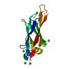
Mass: 40.078 Da / Num. of mol.: 7 / Source method: obtained synthetically / Formula: Ca
#3: Chemical ChemComp-GOL / GLYCEROL / GLYCERIN / PROPANE-1,2,3-TRIOL / Glycerol


Mass: 92.094 Da / Num. of mol.: 1 / Source method: obtained synthetically / Formula: C3H8O3
#4: Water ChemComp-HOH / water / Water


Mass: 18.015 Da / Num. of mol.: 140 / Source method: isolated from a natural source / Formula: H2O

-
Experimental details

-
Experiment

ExperimentMethod: X-RAY DIFFRACTION / Number of used crystals: 1

-
Sample preparation

CrystalDensity Matthews: 1.72 Å3/Da / Density % sol: 28.4 %
Crystal growTemperature: 298 K / Method: evaporation / pH: 8.5
Details: 20% PEG 4000, 0.2M CaCl2, 0.1M Tris-HCl, microbatch, pH 8.5, EVAPORATION, temperature 298.0K

-
Data collection

DiffractionMean temperature: 93 K
Diffraction sourceSource: SYNCHROTRON / Site: APS / Beamline: 23-ID-B / Wavelength: 1.0332
DetectorType: MARMOSAIC 300 mm CCD / Detector: CCD / Date: Dec 6, 2012
RadiationMonochromator: SI(111) / Protocol: SINGLE WAVELENGTH / Monochromatic (M) / Laue (L): M / Scattering type: x-ray
Radiation wavelengthWavelength: 1.0332 Å / Relative weight: 1
ReflectionResolution: 1.35→29.191 Å / Num. obs: 17224 / % possible obs: 95.1 % / Observed criterion σ(I): 0 / Redundancy: 3.6 % / Biso Wilson estimate: 7.4 Å2 / Rmerge(I) obs: 0.049 / Net I/σ(I): 10.33
Reflection shellResolution: 1.35→1.42 Å / Redundancy: 3.2 % / Rmerge(I) obs: 0.085 / Mean I/σ(I) obs: 10.7 / % possible all: 91.6

-
Processing

Software
NameVersionClassification
JBluIce-EPICSdata collection
PHASERphasing
REFMAC5.7.0032refinement
MOSFLMdata reduction
SCALAdata scaling
RefinementMethod to determine structure: MOLECULAR REPLACEMENT
Starting model: PDB ENTRY 4KDV
Resolution: 1.35→29.19 Å / Cor.coef. Fo:Fc: 0.975 / Cor.coef. Fo:Fc free: 0.96 / SU B: 0.657 / SU ML: 0.028 / Cross valid method: THROUGHOUT / ESU R: 0.047 / ESU R Free: 0.053 / Stereochemistry target values: MAXIMUM LIKELIHOOD / Details: HYDROGENS HAVE BEEN ADDED IN THE RIDING POSITIONS
RfactorNum. reflection% reflectionSelection details
Rfree0.165 887 5.1 %RANDOM
Rwork0.13 ---
obs0.132 16337 95.1 %-
all-16337 --
Solvent computationIon probe radii: 0.8 Å / Shrinkage radii: 0.8 Å / VDW probe radii: 1.2 Å / Solvent model: MASK
Displacement parametersBiso mean: 9.71 Å2
Baniso -1Baniso -2Baniso -3
1-0.54 Å20.21 Å20.06 Å2
2--0.03 Å20.21 Å2
3----0.46 Å2
Refinement stepCycle: LAST / Resolution: 1.35→29.19 Å
ProteinNucleic acidLigandSolventTotal
Num. atoms691 0 13 140 844
Refine LS restraints
Refine-IDTypeDev idealDev ideal targetNumber
X-RAY DIFFRACTIONr_bond_refined_d0.0250.02711
X-RAY DIFFRACTIONr_bond_other_d0.0010.02616
X-RAY DIFFRACTIONr_angle_refined_deg2.2441.921983
X-RAY DIFFRACTIONr_angle_other_deg0.92731423
X-RAY DIFFRACTIONr_dihedral_angle_1_deg6.2285105
X-RAY DIFFRACTIONr_dihedral_angle_2_deg27.98928.51927
X-RAY DIFFRACTIONr_dihedral_angle_3_deg8.7041588
X-RAY DIFFRACTIONr_dihedral_angle_4_deg
X-RAY DIFFRACTIONr_chiral_restr0.1280.2133
X-RAY DIFFRACTIONr_gen_planes_refined0.010.02858
X-RAY DIFFRACTIONr_gen_planes_other0.0010.02132
X-RAY DIFFRACTIONr_nbd_refined
X-RAY DIFFRACTIONr_nbd_other
X-RAY DIFFRACTIONr_nbtor_refined
X-RAY DIFFRACTIONr_nbtor_other
X-RAY DIFFRACTIONr_xyhbond_nbd_refined
X-RAY DIFFRACTIONr_xyhbond_nbd_other
X-RAY DIFFRACTIONr_metal_ion_refined
X-RAY DIFFRACTIONr_metal_ion_other
X-RAY DIFFRACTIONr_symmetry_vdw_refined
X-RAY DIFFRACTIONr_symmetry_vdw_other
X-RAY DIFFRACTIONr_symmetry_hbond_refined
X-RAY DIFFRACTIONr_symmetry_hbond_other
X-RAY DIFFRACTIONr_symmetry_metal_ion_refined
X-RAY DIFFRACTIONr_symmetry_metal_ion_other
X-RAY DIFFRACTIONr_mcbond_it
X-RAY DIFFRACTIONr_mcbond_other
X-RAY DIFFRACTIONr_mcangle_it
X-RAY DIFFRACTIONr_mcangle_other
X-RAY DIFFRACTIONr_scbond_it
X-RAY DIFFRACTIONr_scbond_other
X-RAY DIFFRACTIONr_scangle_it
X-RAY DIFFRACTIONr_scangle_other
X-RAY DIFFRACTIONr_long_range_B_refined
X-RAY DIFFRACTIONr_long_range_B_other
X-RAY DIFFRACTIONr_rigid_bond_restr
X-RAY DIFFRACTIONr_sphericity_free
X-RAY DIFFRACTIONr_sphericity_bonded
LS refinement shellResolution: 1.35→1.42 Å / Total num. of bins used: 20
RfactorNum. reflection% reflection
Rfree0.194 --
Rwork0.135 1107 -
obs--90.74 %

+
About Yorodumi

-
News

-
Feb 9, 2022. New format data for meta-information of EMDB entries

New format data for meta-information of EMDB entries

  • Version 3 of the EMDB header file is now the official format.
  • The previous official version 1.9 will be removed from the archive.

Related info.:EMDB header

External links:wwPDB to switch to version 3 of the EMDB data model

-
Aug 12, 2020. Covid-19 info

Covid-19 info

URL: https://pdbj.org/emnavi/covid19.php

New page: Covid-19 featured information page in EM Navigator.

Related info.:Covid-19 info / Mar 5, 2020. Novel coronavirus structure data

+
Mar 5, 2020. Novel coronavirus structure data

Novel coronavirus structure data

Related info.:Yorodumi Speices / Aug 12, 2020. Covid-19 info

External links:COVID-19 featured content - PDBj / Molecule of the Month (242):Coronavirus Proteases

+
Jan 31, 2019. EMDB accession codes are about to change! (news from PDBe EMDB page)

EMDB accession codes are about to change! (news from PDBe EMDB page)

  • The allocation of 4 digits for EMDB accession codes will soon come to an end. Whilst these codes will remain in use, new EMDB accession codes will include an additional digit and will expand incrementally as the available range of codes is exhausted. The current 4-digit format prefixed with “EMD-” (i.e. EMD-XXXX) will advance to a 5-digit format (i.e. EMD-XXXXX), and so on. It is currently estimated that the 4-digit codes will be depleted around Spring 2019, at which point the 5-digit format will come into force.
  • The EM Navigator/Yorodumi systems omit the EMD- prefix.

Related info.:Q: What is EMD? / ID/Accession-code notation in Yorodumi/EM Navigator

External links:EMDB Accession Codes are Changing Soon! / Contact to PDBj

+
Jul 12, 2017. Major update of PDB

Major update of PDB

  • wwPDB released updated PDB data conforming to the new PDBx/mmCIF dictionary.
  • This is a major update changing the version number from 4 to 5, and with Remediation, in which all the entries are updated.
  • In this update, many items about electron microscopy experimental information are reorganized (e.g. em_software).
  • Now, EM Navigator and Yorodumi are based on the updated data.

External links:wwPDB Remediation / Enriched Model Files Conforming to OneDep Data Standards Now Available in the PDB FTP Archive

-
Yorodumi

Thousand views of thousand structures

  • Yorodumi is a browser for structure data from EMDB, PDB, SASBDB, etc.
  • This page is also the successor to EM Navigator detail page, and also detail information page/front-end page for Omokage search.
  • The word "yorodu" (or yorozu) is an old Japanese word meaning "ten thousand". "mi" (miru) is to see.

Related info.:EMDB / PDB / SASBDB / Comparison of 3 databanks / Yorodumi Search / Aug 31, 2016. New EM Navigator & Yorodumi / Yorodumi Papers / Jmol/JSmol / Function and homology information / Changes in new EM Navigator and Yorodumi

Read more