 Movie
Movie Controller

[English] 日本語
 Yorodumi
- PDB-4kdp: TcaR-ssDNA complex crystal structure reveals the novel ssDNA bind... -
+ Open data

- Basic information
| Entry | Database: PDB / ID: 4kdp | ||||||
|---|---|---|---|---|---|---|---|
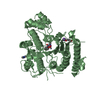
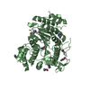
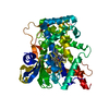
| Title | TcaR-ssDNA complex crystal structure reveals the novel ssDNA binding mechanism of the MarR family proteins | ||||||
 Components Components |
| ||||||
 Keywords Keywords | TRANSCRIPTION/DNA / Multiple drug resistance / ssDNA binding / antibiotics / Staphylococci / TRANSCRIPTION-DNA complex | ||||||
| Function / homology |  Function and homology information Function and homology informationresponse to stress / DNA-binding transcription factor activity / DNA binding Similarity search - Function | ||||||
| Biological species |  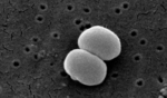 Staphylococcus epidermidis (bacteria) Staphylococcus epidermidis (bacteria) | ||||||
| Method |  X-RAY DIFFRACTION / SYNCHROTRON / MOLECULAR REPLACEMENT / Resolution: 3.6 Å X-RAY DIFFRACTION / SYNCHROTRON / MOLECULAR REPLACEMENT / Resolution: 3.6 Å | ||||||
 Authors Authors | Chang, Y.M. / Chen, C.K.-M. / Wang, A.H.-J. | ||||||
 Citation Citation | Journal: Nucleic Acids Res. / Year: 2014 Title: TcaR-ssDNA complex crystal structure reveals new DNA binding mechanism of the MarR family proteins. Authors: Chang, Y.M. / Ho, C.H. / Chen, C.K. / Maestre-Reyna, M. / Chang-Chien, M.W. / Wang, A.H. | ||||||
| History |
|
- Structure visualization
| Structure viewer | Molecule: MolmilJmol/JSmol |
|---|
- Downloads & links
-Download
| PDBx/mmCIF format | 4kdp.cif.gz | 233 KB | Display | PDBx/mmCIF format |
|---|---|---|---|---|
| PDB format | pdb4kdp.ent.gz | 187.1 KB | Display | PDB format |
| PDBx/mmJSON format | 4kdp.json.gz | Tree view | PDBx/mmJSON format | |
| Others |  Other downloads Other downloads |
-Validation report
| Arichive directory | https://data.pdbj.org/pub/pdb/validation_reports/kd/4kdpftp://data.pdbj.org/pub/pdb/validation_reports/kd/4kdp | HTTPS FTP |
|---|
-Related structure data
| Related structure data |  3kp7S S: Starting model for refinement |
|---|---|
| Similar structure data |


-Links
 PDBj
PDBj


- Assembly
| Deposited unit | 
| ||||||||
|---|---|---|---|---|---|---|---|---|---|
| 1 | 
| ||||||||
| 2 | 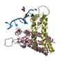
| ||||||||
| 3 | 
| ||||||||
| 4 | 
| ||||||||
| Unit cell |
|
-Components
| #1: Protein | Mass: 17378.195 Da / Num. of mol.: 7 Source method: isolated from a genetically manipulated source Source: (gene. exp.) Staphylococcus epidermidis (bacteria) / Strain: ATCC 12228 / Gene: SE_1937 / Plasmid: pET32 Xa/LIC / Production host: #2: DNA chain | Mass: 5158.339 Da / Num. of mol.: 2 / Source method: obtained synthetically #3: Chemical | ChemComp-EDO /   Mass: 62.068 Da / Num. of mol.: 6 / Source method: obtained synthetically / Formula: C2H6O2 Mass: 62.068 Da / Num. of mol.: 6 / Source method: obtained synthetically / Formula: C2H6O2#4: Chemical | ChemComp-TRS /   Mass: 122.143 Da / Num. of mol.: 5 / Source method: obtained synthetically / Formula: C4H12NO3 / Comment: pH buffer*YM Mass: 122.143 Da / Num. of mol.: 5 / Source method: obtained synthetically / Formula: C4H12NO3 / Comment: pH buffer*YM#5: Water | ChemComp-HOH / |  Mass: 18.015 Da / Num. of mol.: 91 / Source method: isolated from a natural source / Formula: H2O Mass: 18.015 Da / Num. of mol.: 91 / Source method: isolated from a natural source / Formula: H2O |
|---|
-Experimental details
-Experiment
| Experiment | Method: X-RAY DIFFRACTION / Number of used crystals: 1 |
|---|
- Sample preparation
| Crystal | Density Matthews: 2.42 Å3/Da / Density % sol: 49.09 % |
|---|---|
| Crystal grow | Temperature: 298 K / Method: vapor diffusion, sitting drop / pH: 7 Details: 40 % ethylene glycol, 0.1 M Tris , pH 7.0, VAPOR DIFFUSION, SITTING DROP, temperature 298K |
-Data collection
| Diffraction | Mean temperature: 100 K |
|---|---|
| Diffraction source | Source: SYNCHROTRON / Site: SPring-8  / Beamline: BL44XU / Wavelength: 1 Å / Beamline: BL44XU / Wavelength: 1 Å |
| Detector | Type: Bruker DIP-6040 / Detector: CCD / Date: Jan 28, 2012 / Details: mirrors |
| Radiation | Monochromator: GRAPHITE / Protocol: SINGLE WAVELENGTH / Monochromatic (M) / Laue (L): M / Scattering type: x-ray |
| Radiation wavelength | Wavelength: 1 Å / Relative weight: 1 |
| Reflection | Resolution: 3.6→30 Å / Num. all: 15185 / Num. obs: 15179 / % possible obs: 98.8 % / Observed criterion σ(F): 0 / Observed criterion σ(I): 0 / Redundancy: 3 % / Rmerge(I) obs: 0.052 / Net I/σ(I): 18.2 |
| Reflection shell | Resolution: 3.6→3.73 Å / Redundancy: 3 % / Rmerge(I) obs: 0.526 / Mean I/σ(I) obs: 2.1 / % possible all: 99.7 |
- Processing
| Software |
| ||||||||||||||||||||||||||||||||||||||||||||||||||||||||||||
|---|---|---|---|---|---|---|---|---|---|---|---|---|---|---|---|---|---|---|---|---|---|---|---|---|---|---|---|---|---|---|---|---|---|---|---|---|---|---|---|---|---|---|---|---|---|---|---|---|---|---|---|---|---|---|---|---|---|---|---|---|---|
| Refinement | Method to determine structure: MOLECULAR REPLACEMENT Starting model: 3KP7 Resolution: 3.6→16 Å / Isotropic thermal model: isotropic / Cross valid method: THROUGHOUT / σ(F): 0 / Stereochemistry target values: Engh & Huber
| ||||||||||||||||||||||||||||||||||||||||||||||||||||||||||||
| Displacement parameters | Biso mean: 112.238 Å2 | ||||||||||||||||||||||||||||||||||||||||||||||||||||||||||||
| Refinement step | Cycle: LAST / Resolution: 3.6→16 Å
| ||||||||||||||||||||||||||||||||||||||||||||||||||||||||||||
| Refine LS restraints |
| ||||||||||||||||||||||||||||||||||||||||||||||||||||||||||||
| LS refinement shell | Resolution: 3.6→3.73 Å
|
