 Movie
Movie Controller

[English] 日本語
 Yorodumi
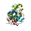
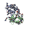
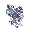
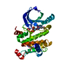
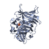
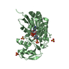
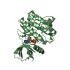
- PDB-4k4a: X-ray crystal structure of E. coli YdiI complexed with phenacyl-CoA -
+ Open data

- Basic information
| Entry | Database: PDB / ID: 4k4a | ||||||
|---|---|---|---|---|---|---|---|
| Title | X-ray crystal structure of E. coli YdiI complexed with phenacyl-CoA | ||||||
 Components Components | Esterase YdiI | ||||||
 Keywords Keywords | HYDROLASE / Hotdog fold / Thioesterase | ||||||
| Function / homology |  Function and homology information Function and homology information1,4-dihydroxy-2-naphthoyl-CoA hydrolase / acyl-CoA hydrolase activity / 1,4-dihydroxy-2-naphthoyl-CoA thioesterase activity / menaquinone biosynthetic process / hydrolase activity / cytosol Similarity search - Function | ||||||
| Biological species |  | ||||||
| Method |  X-RAY DIFFRACTION / MOLECULAR REPLACEMENT / Resolution: 1.89 Å X-RAY DIFFRACTION / MOLECULAR REPLACEMENT / Resolution: 1.89 Å | ||||||
 Authors Authors | Ru, W. / Farelli, J.D. / Dunaway-Mariano, D. / Allen, K.N. | ||||||
 Citation Citation | Journal: Biochemistry / Year: 2014 Title: Structure and Catalysis in the Escherichia coli Hotdog-fold Thioesterase Paralogs YdiI and YbdB. Authors: Wu, R. / Latham, J.A. / Chen, D. / Farelli, J. / Zhao, H. / Matthews, K. / Allen, K.N. / Dunaway-Mariano, D. | ||||||
| History |
|
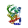
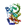
- Structure visualization
| Structure viewer | Molecule: MolmilJmol/JSmol |
|---|
- Downloads & links
-Download
| PDBx/mmCIF format | 4k4a.cif.gz | 133.7 KB | Display | PDBx/mmCIF format |
|---|---|---|---|---|
| PDB format | pdb4k4a.ent.gz | 106.3 KB | Display | PDB format |
| PDBx/mmJSON format | 4k4a.json.gz | Tree view | PDBx/mmJSON format | |
| Others |  Other downloads Other downloads |
-Validation report
| Arichive directory | https://data.pdbj.org/pub/pdb/validation_reports/k4/4k4aftp://data.pdbj.org/pub/pdb/validation_reports/k4/4k4a | HTTPS FTP |
|---|
-Related structure data
| Related structure data | 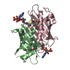 4k49C  4k4bC 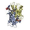 4k4cC 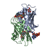 4k4dC  1sbkS S: Starting model for refinement C: citing same article ( |
|---|---|
| Similar structure data |

-Links
 PDBj
PDBj
- Assembly
| Deposited unit | 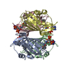
| ||||||||
|---|---|---|---|---|---|---|---|---|---|
| 1 | 
| ||||||||
| 2 | 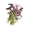
| ||||||||
| Unit cell |
| ||||||||
| Components on special symmetry positions |
|
-Components
| #1: Protein | Mass: 14966.271 Da / Num. of mol.: 4 Source method: isolated from a genetically manipulated source Source: (gene. exp.) References: UniProt: P77781, Hydrolases; Acting on ester bonds #2: Chemical | ChemComp-0FQ / 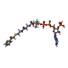  Mass: 885.667 Da / Num. of mol.: 4 / Source method: obtained synthetically / Formula: C29H42N7O17P3S Mass: 885.667 Da / Num. of mol.: 4 / Source method: obtained synthetically / Formula: C29H42N7O17P3S#3: Water | ChemComp-HOH / |  Mass: 18.015 Da / Num. of mol.: 545 / Source method: isolated from a natural source / Formula: H2O Mass: 18.015 Da / Num. of mol.: 545 / Source method: isolated from a natural source / Formula: H2O |
|---|
-Experimental details
-Experiment
| Experiment | Method: X-RAY DIFFRACTION / Number of used crystals: 1 |
|---|
- Sample preparation
| Crystal | Density Matthews: 2.32 Å3/Da / Density % sol: 46.96 % |
|---|---|
| Crystal grow | Temperature: 290 K / Method: vapor diffusion, hanging drop / pH: 6.5 Details: 0.18 M MAGNESIUM ACETATE, 0.07-0.08 M SODIUM CACODYLATE, PH 6.5, VAPOR DIFFUSION, HANGING DROP, temperature 290K |
-Data collection
| Diffraction | Mean temperature: 100 K |
|---|---|
| Diffraction source | Source: ROTATING ANODE / Type: BRUKER AXS MICROSTAR / Wavelength: 1.5418 Å |
| Detector | Type: Bruker Platinum 135 / Detector: CCD / Date: May 9, 2009 |
| Radiation | Monochromator: GRAPHITE / Protocol: SINGLE WAVELENGTH / Monochromatic (M) / Laue (L): M / Scattering type: x-ray |
| Radiation wavelength | Wavelength: 1.5418 Å / Relative weight: 1 |
| Reflection | Resolution: 1.89→44.61 Å / Num. obs: 42793 |
- Processing
| Software |
| ||||||||||||||||||||||||||||||||||||||||||||||||||||||||||||||||||||||||||||||||||||||||||||||||||||||||||||||||
|---|---|---|---|---|---|---|---|---|---|---|---|---|---|---|---|---|---|---|---|---|---|---|---|---|---|---|---|---|---|---|---|---|---|---|---|---|---|---|---|---|---|---|---|---|---|---|---|---|---|---|---|---|---|---|---|---|---|---|---|---|---|---|---|---|---|---|---|---|---|---|---|---|---|---|---|---|---|---|---|---|---|---|---|---|---|---|---|---|---|---|---|---|---|---|---|---|---|---|---|---|---|---|---|---|---|---|---|---|---|---|---|---|---|
| Refinement | Method to determine structure: MOLECULAR REPLACEMENT Starting model: PDB entry 1SBK Resolution: 1.89→44.61 Å / SU ML: 0.16 / σ(F): 1.34 / Phase error: 19.5 / Stereochemistry target values: ML
| ||||||||||||||||||||||||||||||||||||||||||||||||||||||||||||||||||||||||||||||||||||||||||||||||||||||||||||||||
| Solvent computation | Shrinkage radii: 0.9 Å / VDW probe radii: 1.11 Å / Solvent model: FLAT BULK SOLVENT MODEL | ||||||||||||||||||||||||||||||||||||||||||||||||||||||||||||||||||||||||||||||||||||||||||||||||||||||||||||||||
| Refinement step | Cycle: LAST / Resolution: 1.89→44.61 Å
| ||||||||||||||||||||||||||||||||||||||||||||||||||||||||||||||||||||||||||||||||||||||||||||||||||||||||||||||||
| Refine LS restraints |
| ||||||||||||||||||||||||||||||||||||||||||||||||||||||||||||||||||||||||||||||||||||||||||||||||||||||||||||||||
| LS refinement shell |
|
