Mass: 18.015 Da / Num. of mol.: 74 / Source method: isolated from a natural source / Formula: H2O
Has protein modification
Y
-
Experimental details
-
Experiment
Experiment
Method: X-RAY DIFFRACTION / Number of used crystals: 1
-
Sample preparation
Crystal
Density Matthews: 3.64 Å3/Da / Density % sol: 66.21 %
Crystal grow
Temperature: 292 K / Method: vapor diffusion / pH: 4.2 Details: 100 mM Na citrate, 33% PEG400 and 200 mM LiSO4, VAPOR DIFFUSION, temperature 292K, pH 4.2
Resolution: 2.1→2.21 Å / Redundancy: 6.2 % / Mean I/σ(I) obs: 3.99 / Rsym value: 0.424 / % possible all: 99.7
-
Processing
Software
Name
Version
Classification
XDS
datascaling
PHASER
phasing
REFMAC
5.7.0029
refinement
XDS
datareduction
XSCALE
datascaling
Refinement
Method to determine structure: SAD / Resolution: 2.1→28.57 Å / Cor.coef. Fo:Fc: 0.959 / Cor.coef. Fo:Fc free: 0.963 / SU B: 2.19 / SU ML: 0.062 / Cross valid method: THROUGHOUT / ESU R: 0.023 / ESU R Free: 0.021 / Stereochemistry target values: MAXIMUM LIKELIHOOD / Details: HYDROGENS HAVE BEEN USED IF PRESENT IN THE INPUT
Rfactor
Num. reflection
% reflection
Selection details
Rfree
0.1802
563
4.1 %
RANDOM
Rwork
0.16239
-
-
-
obs
0.16313
13261
99.42 %
-
all
-
13291
-
-
Solvent computation
Ion probe radii: 0.8 Å / Shrinkage radii: 0.8 Å / VDW probe radii: 1.2 Å / Solvent model: MASK
 Movie
Movie Controller


 Open data
 Basic information
 Components
 Keywords
 Function and homology information
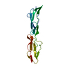
 Homo sapiens (human)
 X-RAY DIFFRACTION /
  Authors
 Citation
 Structure visualization
 Downloads & links
 Other downloads

 PDBj
PDBj

 Assembly


 Mass: 194.226 Da / Num. of mol.: 2 / Source method: obtained synthetically / Formula: C8H18O5 / Comment: precipitant*YM
 Mass: 96.063 Da / Num. of mol.: 1 / Source method: obtained synthetically / Formula: SO4
 Mass: 18.015 Da / Num. of mol.: 74 / Source method: isolated from a natural source / Formula: H2O
 Sample preparation
 / Beamline: I02 / Wavelength: 0.978 Å
 Processing